38 draw and label the parts of the microscope
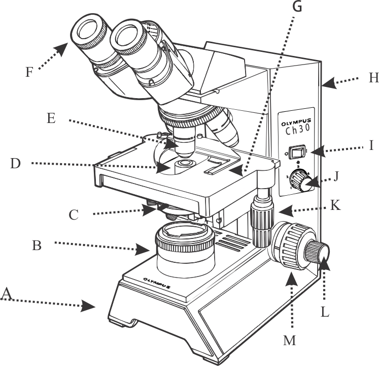
PDF Parts of the compound microscope: Write the correct label for each part ... Draw this table, and then place a check mark or "X" indicating a structure/organelle is present in the indicated cell type. Cell Type Nucleus Endoplasmic Reticulum Golgi Apparatus Mitochondrion Chloroplast Plasma Membrane Cell Wall Flagellum Bacteria Plant Animal 22 23 24 25 26 27 28 29 30 32 31 33 34 35 PDF Label parts of the Microscope: Answers Label parts of the Microscope: Answers Coarse Focus Fine Focus Eyepiece Arm Rack Stop Stage Clip . Created Date: 20150715115425Z ...
Labeling the Parts of the Microscope | Microscope activity, Science ... Description A collection of microscope diagrams and worksheets for science class. Download them all in one convenient PDF, and select the version that's best for your classroom. This PDF contains the following: 1. Parts of a Microscope Diagram - Color 2. Parts of a Microscope Diagram - Black and White 3.

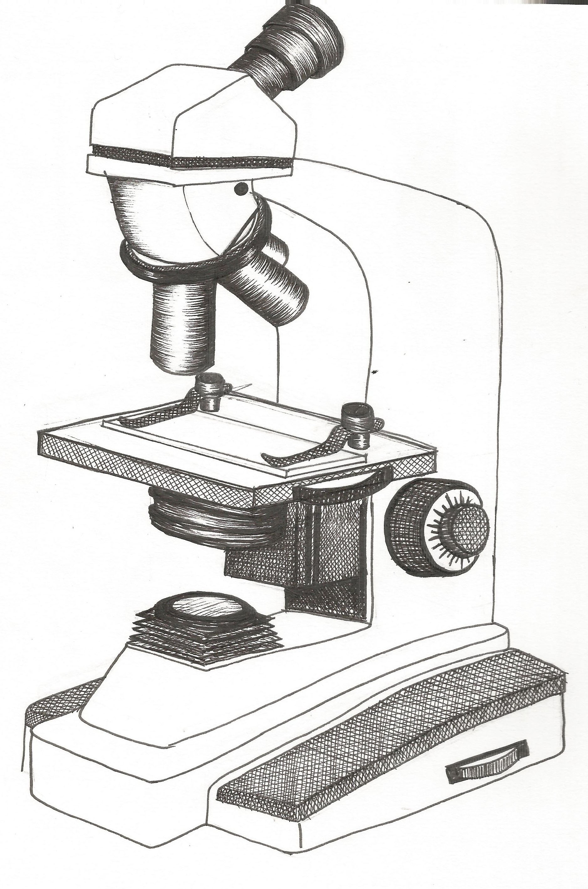
Draw and label the parts of the microscope
Microscope Parts Drawing - Painting Valley All the best Microscope Parts Drawing 36+ collected on this page. Feel free to explore, study and enjoy paintings with PaintingValley.com. ... Solved Label The Par... 323x252 0 0. Like JPG. The Microscope - Mic... 450x474 0 0. Like PNG. What Is A Microscope... 888x762 0 0. Like JPG. Exe - Microscope Par... 933x1163 0 0. PDF Label and Color the Microscope Parts - White Plains Public Schools Label and Color the Microscope Parts Color the arm dark green Color the base red Color the stage dark blue. Draw orange stripes on the body tube Color the diaphragm light green. Color the eyepiece black. Color the stage clips purple. Color the nosepiece light blue. Color the light source yellow Draw green stripes on the inclination joint Microscope, Microscope Parts, Labeled Diagram, and Functions The description given below summarize the brief description of microscope parts used to visualize the microscopic specimens such as animal cells, plant cells, microbes, bacteria, viruses, microorganisms etc. The Microscopes parts divided into three different structural parts Head, Base, and Arms.
Draw and label the parts of the microscope. Label the parts of a microscope ~(˘ ˘~) - Brainly.com (~˘ ˘)~ Label the parts of a microscope ~(˘ ˘~) - 16653121 xXPhantomWarriorXx xXPhantomWarriorXx 05/28/2020 Biology High School answered (~˘ ˘)~ Label the parts of a microscope ~(˘ ˘~) 1 See answer Advertisement Advertisement xXPhantomWarriorXx is waiting for your help. Add your answer and earn points. Parts of the Microscope Printables - ThoughtCo Most microscopes used in a classroom setting are compound microscopes. These usually consist of a light source and three to five lenses with a total magnification of 40x to 1000x. The following free printables can help you teach your students the basic parts of a microscope so that they are ready to dive into a world previously unseen. A Study of the Microscope and its Functions With a Labeled Diagram These labeled microscope diagrams and the functions of its various parts, attempt to simplify the microscope for you. However, as the saying goes, 'practice makes perfect', here is a blank compound microscope diagram and blank electron microscope diagram to label. Download the diagrams and practice labeling the different parts of these ... Simple Microscope - Parts, Functions, Diagram and Labelling Stage - The stage of the microscope is a metal plate that is rectangular in shape and fitted to the vertical rod. It comes with a hole in the center that enables the light to pass from below. The stage holds the slide that contains the specimen to be examined for.
Directions: Draw and label the parts of the microscope.(Name) Directions: Draw and label the parts of the microscope.(Name) - 7174938 mjcray43 mjcray43 19.11.2020 Integrated Science Junior High School answered Directions: Draw and label the parts of the microscope.(Name) 1 See answer hi alam mo sagot geh pashneya gg Advertisement Advertisement Parts of a microscope with functions and labeled diagram Q. List down the 18 parts of a Microscope. 1. Ocular Lens (Eye Piece) 2. Diopter Adjustment 3. Head 4. Nose Piece 5. Objective Lens 6. Arm (Carrying Handle) 7. Mechanical Stage 8. Stage Clip 9. Aperture 10. Diaphragm 11. Condenser 12. Coarse Adjustment 13. Fine Adjustment 14. Illuminator (Light Source) 15. Stage Controls 16. Base 17. Draw and label the parts of the microscope. Draw it in your science ... Draw and label the parts of the microscope. Draw it in your science activity notebook and label each part numbered 1-14. 1 See answer Thank you mali Po yan Advertisement jackelynbetonio Answer: 1. Body tube 2. Arm 3. HPO / High Power Objective 4. Oil Immersion 5. LPO / Low Power Objective 6. Stage clip 7. Base 8. Inclination joint 9. Mirror 10. Parts of Microscope, Function, Names & Labeled Diagram - slidingmotion Microscope parts labeled diagram gives us all the information about its parts and their position in the microscope. Microscope Parts Labeled Diagram The principle of the Microscope gives you an exact reason to use it. It works on the 3 principles. Magnification Resolving Power Numerical Aperture. Parts of Microscope Head Base Arm Eyepiece Lens
Labeling the Parts of the Microscope Labeling the Parts of the Microscope This activity has been designed for use in homes and schools. Each microscope layout (both blank and the version with answers) are available as PDF downloads. You can view a more in-depth review of each part of the microscope here. Download the Label the Parts of the Microscope PDF printable version here. Compound Microscope Parts - Labeled Diagram and their Functions - Rs ... There are three major structural parts of a compound microscope. The head includes the upper part of the microscope, which houses the most critical optical components, and the eyepiece tube of the microscope. The base acts as the foundation of microscopes and houses the illuminator. The arm connects between the base and the head parts. Compound Microscope Parts, Functions, and Labeled Diagram Compound Microscope Definitions for Labels. Eyepiece (ocular lens) with or without Pointer: The part that is looked through at the top of the compound microscope. Eyepieces typically have a magnification between 5x & 30x. Monocular or Binocular Head: Structural support that holds & connects the eyepieces to the objective lenses. Parts Of The Microscope Label Worksheets & Teaching Resources | TpT Learn the parts of a microscope with this resource!Included in this resource are two pdf documents. One is an answer key/review sheet of a labeled microscope. The other is the microscope with the label boxes blank that I used as the 'quiz'. (Files include a link to editable doc, so you can rewrite a
NCERT Exemplar Problems Class 9 Science - The Fundamental Unit of Life ...
Color the Parts of the Microscope - The Biology Corner Light microscopes use either a bulb or a mirror (M) as their light source. Color the light source yellow . The switch for this light is usually found on the base of the microscope, and sometimes on the power cord. You can control how much light goes through the specimen by adjusting the diaphragm (K). Color the diaphragm light green .

The Fundamental Unit of Life : NCERT Exemplar - Page 3 of 3 - DronStudy.com
PDF Parts of a Microscope Printables - Homeschool Creations Parts of a eyepiece arm stageclips nosepiece focusing knobs illuminator stage objective lenses head base Label the parts of the microscope. You can use the word bank below to fill in the blanks or cut and paste the words at the bottom. Microscope Created by Jolanthe @ HomeschoolCreations.net eyepiece head objective lenses arm focusing knob base ...

Integumentary system anatomy and physiology (skin anatomy ...
Microscope Parts & Functions - AmScope Total magnification of a microscope is determined by the sum of the eyepiece magnification multiplied by that of the objective lens. Eyepiece Tube: The tube in which the eyepiece lens is situated. Fine Focus: A knob used to fine-tune the focus of a specimen in conjunction with the coarse focus.

DRAW IT NEAT : How to draw Chlamydomonas
Label the microscope — Science Learning Hub Use this interactive to identify and label the main parts of a microscope. Drag and drop the text labels onto the microscope diagram. eye piece lens coarse focus adjustment high-power objective diaphragm or iris base fine focus adjustment light source stage Download Exercise Tweet
Compound Microscope Drawing at PaintingValley.com | Explore collection ...
How to draw and label the parts of a microscope? What are at least 3 ... Answer (1 of 3): Google images of light microscope, phase contrast microscope, fluorescent microscope, transmission electron microscope, scanning electron microscope. Label eyepiece, phototube, objectives, stage, sub stage condenser, light (electron) source, focusing knobs, and microscope base. ...

Chapter 1: What is Science - mrs.bagwell.biology
Parts of the Microscope with Labeling (also Free Printouts) Parts of the Microscope with Labeling (also Free Printouts) A microscope is one of the invaluable tools in the laboratory setting. It is used to observe things that cannot be seen by the naked eye. Table of Contents 1. Eyepiece 2. Body tube/Head 3. Turret/Nose piece 4. Objective lenses 5. Knobs (fine and coarse) 6. Stage and stage clips 7. Aperture
Free Microscope Drawing, Download Free Microscope Drawing png images ...
Microscope Parts and Functions All of the parts of a microscope work together - The light from the illuminator passes through the aperture, through the slide, and through the objective lens, where the image of the specimen is magnified.
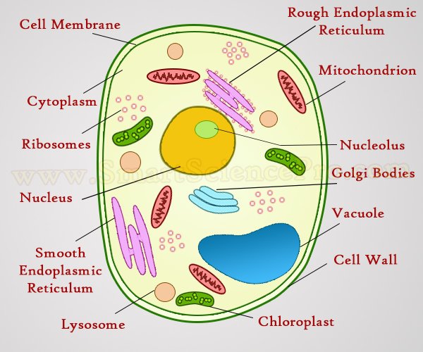
Structure of Animal Cell and Plant Cell Under Microscope
16 Parts of a Compound Microscope: Diagrams and Video Once you have an understanding of the parts of the microscope it will be much easier to navigate around and begin observing your specimen, which is the fun part! The 16 core parts of a compound microscope are: Head (Body) Arm Base Eyepiece Eyepiece tube Objective lenses Revolving Nosepiece (Turret) Rack stop Coarse adjustment knobs
Post a Comment for "38 draw and label the parts of the microscope"