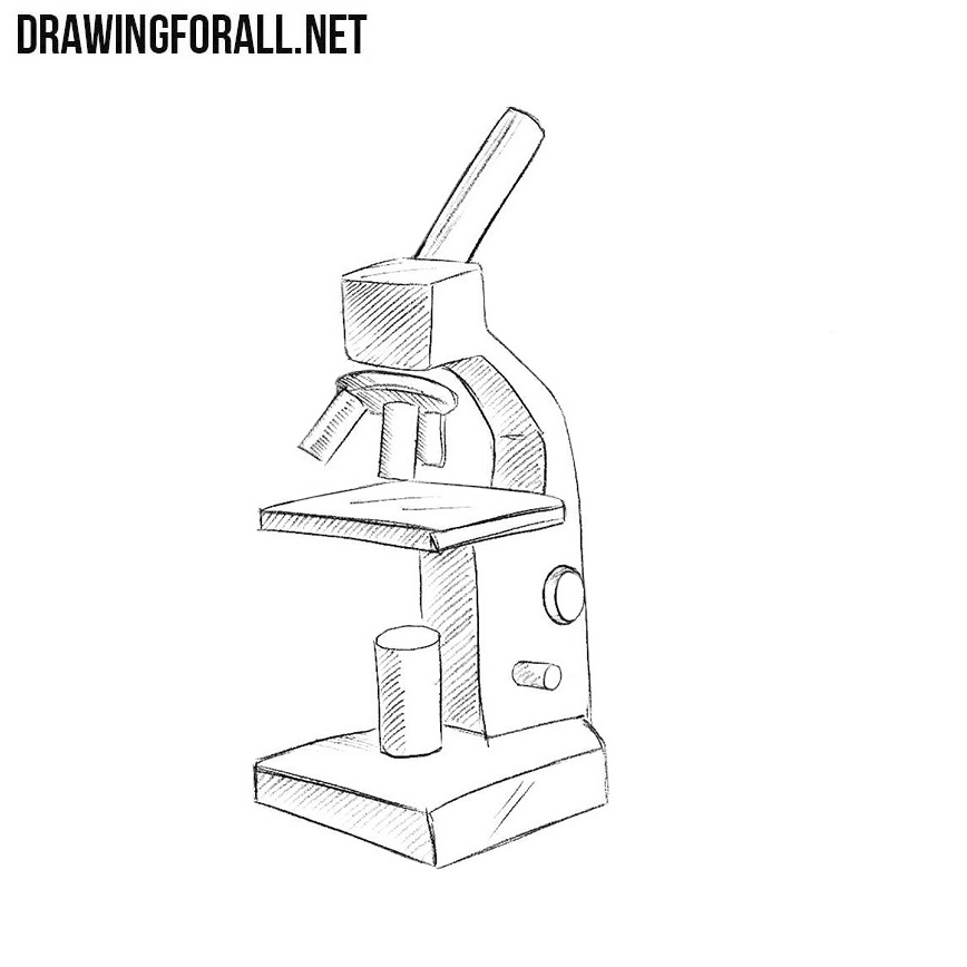
44 drawing of microscope and label
Compound Microscope Parts - Labeled Diagram and their Functions - Rs ... Two adjustment knobs are used to focus the microscope: fine focus knob and coarse focus knob. Both knobs can move the stage up and down. You should use the coarse focus knob to bring the specimen into approximate or near focus. Then you use the fine focus knob to sharpen the focus quality of the image. How to Sketch a Microscope Slide - Identifying and Sketching Cell ... First, to represent the microscope field of view, draw a circle on the page - this can be freehand or, if you want to be precise, use a compass. If you are using a graticule slide (a microscope slide with millimeter grid lines), lightly sketch a grid over your circle.
Microscope drawing Images, Stock Photos & Vectors | Shutterstock Microscope drawing royalty-free images 18,577 microscope drawing stock photos, vectors, and illustrations are available royalty-free. See microscope drawing stock video clips Image type Orientation Color People Artists More Sort by Popular Science Abstract Designs and Shapes College and University Art Styles Printing, Typography, and Calligraphy

Drawing of microscope and label
Parts of a microscope with functions and labeled diagram Figure: Diagram of parts of a microscope There are three structural parts of the microscope i.e. head, base, and arm. Head - This is also known as the body. It carries the optical parts in the upper part of the microscope. Base - It acts as microscopes support. It also carries microscopic illuminators. Microscope Parts and Functions Most specimens are mounted on slides, flat rectangles of thin glass. The specimen is placed on the glass and a cover slip is placed over the specimen. This allows the slide to be easily inserted or removed from the microscope. It also allows the specimen to be labeled, transported, and stored without damage. Microscope, Microscope Parts, Labeled Diagram, and Functions Microscope, Microscope Parts, Labeled Diagram, and Functions What is Microscope? A microscope is a laboratory instrument used to examine objects that are too small to be seen by the naked eye. It is derived from Ancient Greek words and composed of mikrós, "small" and skopeîn,"to look" or "see".
Drawing of microscope and label. PDF Label parts of the Microscope: Answers Label parts of the Microscope: Answers Coarse Focus Fine Focus Eyepiece Arm Rack Stop Stage Clip . Created Date: 20150715115425Z ... Microscope Drawing Easy with Label - YouTube In this video I go over a microscope drawing that is easy with label. There is a blank copy at the end of the video to review on your own. A great way to s... Microscope labeled diagram - SlideShare Microscope labeled diagram 1. The Microscope Image courtesy of: Microscopehelp.com Basic rules to using the microscope 1. You should always carry a microscope with two hands, one on the arm and the other under the base. 2. You should always start on the lowest power objective lens and should always leave the microscope on the low power lens ... learning-center.homesciencetools.com › articleMicroscope Lab Experiments: An Introduction to the Microscope Microscope Worksheet: How to Record Microscope Observations In the field of science, recording observations while performing an experiment is one of the most useful tools available. Early scientists often kept very detailed journals of the experiments they performed, making entries for each individual experiment and writing down virtually ...
Labeling the Parts of the Microscope | Microscope activity, Science ... Description Worksheet identifying the parts of the compound light microscope. Answer key: 1. Body tube 2. Revolving nosepiece 3. Low power objective 4. Medium power objective 5. High power objective 6. Stage clips 7. Diaphragm 8. Light source 9. Eyepiece 10. Arm 11. Stage 12. Coarse adjustment knob 13. Fine adjustment knob 14. Base S › products › microscopeMicroscope Imaging Software | Products | Leica Microsystems Jun 15, 2021 · A range of specific modules allows configuration of the microscope as a dedicated high-performance tool for almost any application. The latest software platform, LAS X, encompasses all microscope solutions for Life Science and Industry applications, offering maximum flexibility. The previous Leica Application Suite continues to be supported. microscope with labels - Openclipart microscope with labels by johnny_automatic - uploaded on October 21, 2006, 11:54 am a side view drawing of a microscope with parts labeled. From "The Brain in Space" produced by NASA and stated as PD. 1 Tags equipment instrument lab laboratory microscope nasa Safe for Work? Yes Download SVG (Vector) PNG (Bitmap) Small Medium Large Microscope Drawing: How to Sketch Microscope Slides How to Draw Microscope Slides Organize and orient your field of view: To begin, draw a circle as large as possible with a pencil. An 8.5 x 11-inch piece of paper is good size for beginners. The circle represents what you see through the eyepiece of the microscope. Using thin lines, divide the circle into quarters in order to organize the picture.
Microscope Drawing And Label - Painting Valley We collected 33+ Microscope Drawing And Label paintings in our online museum of paintings - PaintingValley.com. ADVERTISEMENT LIMITED OFFER: Get 10 free Shutterstock images - PICK10FREE label microscope diagram compound parts light labeling functions microscopic blank labeled biology microscopy labelled beautiful Compound Microscope ... Parts of the Microscope with Labeling (also Free Printouts) Parts of the Microscope with Labeling (also Free Printouts) A microscope is one of the invaluable tools in the laboratory setting. It is used to observe things that cannot be seen by the naked eye. Table of Contents 1. Eyepiece 2. Body tube/Head 3. Turret/Nose piece 4. Objective lenses 5. Knobs (fine and coarse) 6. Stage and stage clips 7. Aperture Microscope Drawing Teaching Resources | Teachers Pay Teachers This GEEKS lab sheet gives students room to draw three specimens. It includes labels for the Specimen Name, Magnification, Observations/ Challenges. ... it shows student work samples (compares A+ work to B and C work) of slide drawings. This will model microscope slide drawings so your students understand the level of detail that is desired ... Microscope Labeling - The Biology Corner Students label the parts of the microscope in this photo of a basic laboratory light microscope. Can be used for practice or as a quiz. ... Microscope Labeling . Microscope Use: 15. When focusing a specimen, you should always start with the _____ objective. 16. When using the high power objective, only the _____ knob should be used. 17. The ...
Histology Slides Database: histological diagram of transverse section ...
A Study of the Microscope and its Functions With a Labeled Diagram ... sciencestruck.com A Study of the Microscope and its Functions With a Labeled Diagram To better understand the structure and function of a microscope, we need to take a look at the labeled microscope diagrams of the compound and electron microscope. These diagrams clearly explain the functioning of the microscopes along with their respective parts.

Rebekah's Dissection Log: Hydra: Phylum Cnideria
Compound Microscope - Diagram (Parts labelled), Principle and Uses The three structural components include: 1. Head - This is the upper part of the microscope that houses the optical parts 2. Arm - This part connects the head with the base and provides stability to the microscope. Arm is used to carry the microscope around 3. Base - Base is on which the microscope rests and the base houses the illuminator that lights up the specimens

Image result for dicot root diagram | Biology diagrams, Root diagram ...
Label Microscope Diagram - EnchantedLearning.com Using the terms listed below, label the microscope diagram. arm - this attaches the eyepiece and body tube to the base. base - this supports the microscope. body tube - the tube that supports the eyepiece. coarse focus adjustment - a knob that makes large adjustments to the focus. diaphragm - an adjustable opening under the stage, allowing ...
4-Color Figures | Engineering360
› iet › microscopeVirtual Microscope - NCBioNetwork.org Lesson Description BioNetwork’s Virtual Microscope is the first fully interactive 3D scope - it’s a great practice tool to prepare you for working in a science lab. Explore topics on usage, care, terminology and then interact with a fully functional, virtual microscope.
How to Draw a Microscope Easy
The Parts of a Microscope (Labeled) Printable - TeacherVision The Parts of a Microscope (Labeled) Printable. Download. Add to Favorites. Share. This diagram labels and explains the function of each part of a microscope. Use this printable as a handout or transparency to help prepare students for working with laboratory equipment. Grade:

White Blood Cells Diagram
PDF Parts of a Microscope Printables - Homeschool Creations Label the parts of the microscope. You can use the word bank below to fill in the blanks or cut and paste the words at the bottom. Microscope Created by Jolanthe @ HomeschoolCreations.net. Parts of a eyepiece arm stageclips nosepiece focusing knobs illuminator stage objective lenses

Microscope Clip Art at Clker.com - vector clip art online, royalty free ...
Microscope Illustrations and Stock Art. 68,272 Microscope illustration ... Stock Illustrations by Morphart 7 / 971 Black microscope Drawings by docent 5 / 1,432 microscope vector Drawing by yupiramos 6 / 694 Microscope sketch Stock Illustration by lhfgraphics 5 / 2,930 Microscope Clip Art by paulfleet 5 / 880 Microscope Stock Illustrations by krishnacreation 1 / 167 Germs under the microscope.

Microscope | ClipArt ETC
Labeling the Parts of the Microscope Labeling the Parts of the Microscope This activity has been designed for use in homes and schools. Each microscope layout (both blank and the version with answers) are available as PDF downloads. You can view a more in-depth review of each part of the microscope here. Download the Label the Parts of the Microscope PDF printable version here.
Exploration of Human Brain Tissue
Label the microscope — Science Learning Hub In this interactive, you can label the different parts of a microscope. Use this with the Microscope parts activity to help students identify and label the main parts of a microscope and then describe their functions. Drag and drop the text labels onto the microscope diagram.
Post a Comment for "44 drawing of microscope and label"