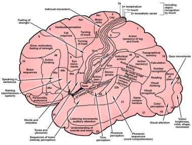
43 correctly label the following functional regions of the cerebral cortex.
› 36111379 › DiFiores_Atlas_ofDiFiore's Atlas of Histology with Functional ... - Academia.edu Enter the email address you signed up with and we'll email you a reset link. › pmc › articlesSARS-CoV-2 is associated with changes in brain structure in ... Mar 07, 2022 · In fact, in a recent functional connectivity study of the primary olfactory cortex, the orbitofrontal cortex was found to be connected to all four primary olfactory regions investigated (frontal and temporal piriform cortex, anterior olfactory nucleus and olfactory tubercle), possibly explaining why it is reliably activated even in basic and ...
Solved Correctly label the following functional regions of - chegg.com Expert Answer 100% (20 ratings) Transcribed image text: Correctly label the following functional regions of the cerebral cortex. Auditory association Wernicke area Visual association area area Primary gustatory cortex Primary auditory cortex Primary visual cortex Previous question Next question

Correctly label the following functional regions of the cerebral cortex.
Solved Correctly label the following functional regions of - Chegg Question: Correctly label the following functional regions of the cerebral cortex Somesthetic association Primary somesthetic cortex area Broca area Prefrontal cortex Primary motor cortex Ofactory association are Motor association area Wh 110 NE 9 o 4 Q W E R S D F G H J K This problem has been solved! See the answer Show transcribed image text Solved Correctly label the following functional regions of - chegg.com Expert Answer 100% (9 ratings) Transcribed image text: Correctly label the following functional regions of the cerebral cortex Somesthetic association area Primary somesthetic cortex Broca area Olfactory association area Motor association area Primary motor cortex Prefrontal cortex Previous question Next question Chapter 14 Flashcards | Quizlet Thalamus Epithalamus Hypothalamus Correctly label the following functional regions of the cerebral cortex. T/F A lesion in the right side of the brainstem will usually cause a sensory or motor deficit on left side of the head. false since fibres cross at the level of thalamus below the level of thalamus , the affect will be seen in same side .
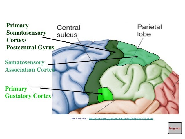
Correctly label the following functional regions of the cerebral cortex.. Solved Correctly label the following functional regions of - chegg.com Question:Correctly label the following functional regions of the cerebral cortex. Auditory association Primary auditory cortex area Primary gustatory cortex Primary visual cortex Visual association area Wernicke area This problem has been solved! See the answerSee the answerSee the answerdone loading Show transcribed image text Expert Answer Brain & CN Worksheet Flashcards - Quizlet Correctly label the following functional regions of the cerebral cortex. Consider a situation where a stroke or mechanical trauma has occurred resulting in damage to one of the areas of the brain indicated in the image. Drag each label into the proper location in order to identify the area that would most likely have been affected. Solved Correctly label the following functional regions of - Chegg Question: Correctly label the following functional regions of the cerebral cortex. Primary motor cortex Primary olfactory cortex Primary somatosensory cortex Primary visual cortex Primary gustatory cortex Primary auditory cortex This problem has been solved! See the answer Show transcribed image text Expert Answer 100% (10 ratings) Chapter 13 Question Set Flashcards - Quizlet Correctly label the following functional regions of the cerebral cortex. Label the regions involved in interpreting and carrying out speech information. Label the diagram with the terms provided to describe the process of neurulation. Cerebrospinal fluid enters the third ventricle of the brain by way of the interventricular foramina.
CBIO Figures Flashcards | Quizlet Correctly label the following functional regions of the cerebral cortex. Consider a situation where a stroke or mechanical trauma has occurred resulting in damage to one of the areas of the brain indicated in the image. Drag each label into the proper location in order to identify the area that would most likely have been affected. Chapter 14 Worksheet Flashcards - Quizlet Correctly label the following functional regions of the cerebral cortex. Consider a situation where a stroke or mechanical trauma has occurred resulting in damage to one of the areas of the brain indicated in the image. Drag each label into the proper location in order to identify the area that would most likely have been affected. Solved Correctly label the following functional regions of - chegg.com Question: Correctly label the following functional regions of the cerebral cortex. Primary auditory cortex Auditory association area Wernicke area Visual association area Primary gustatory cortex Primary visual cortex -ces < Prev 13 of 15 Next > This problem has been solved! See the answer Show transcribed image text Expert Answer 100% (6 ratings) Cerebral Cortex | Facts, Layers, Levels, Functions & Summary The cerebral cortex can be divided into three basic levels and functions: primary secondary tertiary cortex The hierarchically lowest areas are the primary visual, auditory, somatosensory, and motor cortex. The primary sensory cortex receives information through the thalamus. Primary cortex
› tutorial › human_ecogAnalysis of human ECoG and sEEG recordings - FieldTrip toolbox Apr 15, 2022 · Preprocessing of the anatomical MRI. 2) Import the anatomical MRI into the MATLAB workspace using ft_read_mri.The MRI comes in the format of a single file with an .img or .nii extension, or a folder containing a series of files with a .dcm or .ima extension (DICOM; Supplementary File 2 of the original paper may aid in the search and visualization of a DICOM series). The brain Flashcards | Quizlet One of the last areas of the brain to mature fully is the prefrontal cortex, which controls logic, judgment, and expression of emotion. Acting mostly as a communication relay center due to the many fiber tracts through the area, the midbrain includes the cerebral peduncles, colliculi, and nuclei of cranial nerves III and IV. Functions of the Cerebral Cortex - Bodytomy Location of the Cerebral Cortex The outer region of the cerebrum is the cerebral cortex. It is about 1.5mm to 5mm in thickness. The cerebral cortex consists of four lobes; frontal lobe, parietal lobe, temporal lobe, and occipital lobe. The cerebral cortex is a highly convoluted or folded outer layer of the cerebrum. academic.oup.com › brain › articleExpression of 4E-BP1 in juvenile mice alleviates mTOR-induced ... There are four HCN isoforms in the brain; HCN1 and HCN2 are highly expressed in the cortex and hippocampus, whereas HCN3 and HCN4 are predominantly found in the subcortical regions. 42, 43 Within the cortex, HCN1 and 2 are expressed in the deeper layer pyramidal neurons, while no functional HCN channels are found in L2/3 pyramidal neurons. 41 ...
32 Correctly Label The Following Functional Regions Of The Cerebral ...
› pmc › articlesImaging of Intracranial Hemorrhage - PMC Dec 12, 2016 · Non-traumatic IPH centered in the cerebral cortex should prompt consideration of diagnoses other than hypertension, as described in below. Similarly, IPH in patients younger than 50 should prompt consideration of other causes of bleeding such as an underlying brain neoplasm or vascular malformation.
Tayloredge - Science
› 41956428 › COGNITIVE_NEUROSCIENCECOGNITIVE NEUROSCIENCE THE BIOLOGY OF THE MIND Fourth Edition Enter the email address you signed up with and we'll email you a reset link.

35 Correctly Label The Following Anatomical Features Of The Cerebellum ...
Correctly Label The Following Functional Regions Of The Cerebral Cortex ... Correctly Label The Following Functional Regions Of The Cerebral Cortex Somesthetic Association Area Primary Somesthetic Cortex Broca Area Olfactory Association Area Motor Association Area Primary Motor Cortex Prefrontal Cortex Mar 29 2022 10:16 AM Expert's Answer Solution.pdf Next Previous
Brain Anatomy: Overview, Gross Anatomy: Cerebrum, Gross Anatomy: Cortex
Solved Chapter 14 Homework Interactive Questi... Saved Help - chegg.com Saved Help Save& Exit Submit 4 Correctly label the following functional regions of the cerebral cortex. Broca area Prefrontal cortex Olfactory association area Somesthetic association area Primary somesthetic cortexMotor association area 1.5 points Primary motor cortex eBook References < Prev 4of 20 Next >
Brain cortical regions and functions
Critical Thinking 5 Flashcards & Practice Test - Quizlet Correctly label the following functional regions of the cerebral cortex. Primary visual cortex, Primary auditory cortex, Primary gustatory cortex, Primary motor cortex, Primary somatosensory cortex, & Primary olfactory cortex What structure is essential in storing memories and forming long-term memory? hippocampus
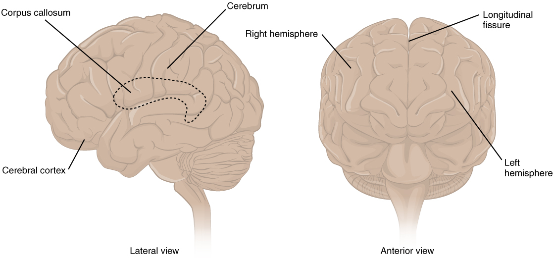
This figure shows the lateral view on the left panel and anterior view ...
Chapter 13 QS Anatomy (Brain and Cranial Nerves) Flashcards - Quizlet Match the function to the correct lobe of the cerebral cortex. 1. Voluntary skeletal muscle control, verbal communication=frontal lobe 2. Auditory association area=temporal lobe 3. Primary gustatory cortex=insular lobe 4. Somatosensory cortex, somatosensory association area=parietal lobe 5. Primary visual cortex=occipital lobe
Post a Comment for "43 correctly label the following functional regions of the cerebral cortex."