43 microscope drawing with label
Collection of Microscope Drawing (32) - Clipart Library Satin Stitch Embroidery Design: Microscope Outline 3.50 inches H x. easy drawing of microscope. Label Microscope Diagram. electron microscope to label. 18,634 Microscope drawing Images, Stock Photos & Vectors - Shutterstock Find Microscope drawing stock images in HD and millions of other royalty-free stock photos, illustrations and vectors in the Shutterstock collection. Thousands of new, high-quality pictures added every day.
Microscope Drawing Easy with Label - YouTube In this video I go over a microscope drawing that is easy with label. There is a blank copy at the end of the video to review on your own. A great way to s...
Microscope drawing with label
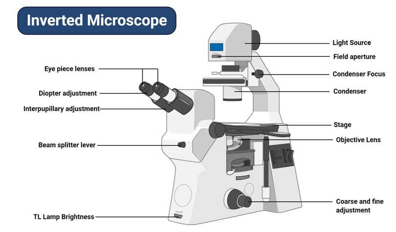
A Study of the Microscope and its Functions With a Labeled Diagram A Study of the Microscope and its Functions With a Labeled Diagram To better understand the structure and function of a microscope, we need to take a look at the labeled microscope diagrams of the compound and electron microscope. These diagrams clearly explain the functioning of the microscopes along with their respective parts. Microscope Drawing: How to Sketch Microscope Slides How to Draw Microscope Slides Organize and orient your field of view: To begin, draw a circle as large as possible with a pencil. An 8.5 x 11-inch piece of paper is good size for beginners. The circle represents what you see through the eyepiece of the microscope. Using thin lines, divide the circle into quarters in order to organize the picture. Compound Microscope- Definition, Labeled Diagram, Principle, Parts, Uses The optical microscope often referred to as the light microscope, is a type of microscope that uses visible light and a system of lenses to magnify images of small subjects. There are two basic types of optical microscopes: Simple microscopes. Compound microscopes. The term "compound" in compound microscopes refers to the microscope having ...
Microscope drawing with label. How To Draw A Microscope - YouTube Today, we're learning how to draw a cool microscope!👩🎨 JOIN OUR ART HUB MEMBERSHIP! VISIT 🎨 VISIT OUR AMAZON ART SUPPLY S... Microscope Diagram and Functions | Science fair projects, Microscope ... A Study of the Microscope and its Functions With a Labeled Diagram To better understand the structure and function of a microscope, we need to take a look at the labeled microscope diagrams of the compound and electron microscope. These diagrams clearly explain the functioning of the microscopes along with their respective parts. M mooketsi Microscope Labeling Diagram | Quizlet PGFry210. Unit 2 Lesson 5 - Punnett Squares and Pedigrees. 4 terms. PGFry210. Unit 2 Lesson 4 - Heredity. 9 terms. PGFry210. Upgrade to remove ads. Only $2.99/month. Compound Microscope - Diagram (Parts labelled), Principle and Uses Image : Labeled Diagram of compound microscope parts. See: Labeled Diagram showing differences between compound and simple microscope parts Structural Components. The three structural components include. 1. Head. This is the upper part of the microscope that houses the optical parts. 2. Arm . This part connects the head with the base and ...
DOC Microscope drawings - University of Manitoba 3. Finish drawing the object or specimen following the same procedure as outlined in steps 1 to 5 of the macroscopic drawings. Remember the rules for good scientific drawings: 1. Use a sharp F, H or 2H pencil for both drawings and labels. 2. Use unlined paper only. 3. Draw clearly defined structures with smooth, continuous lines. 4. Compound Microscope Parts - Labeled Diagram and their Functions - Rs ... Labeled diagram of a compound microscope Major structural parts of a compound microscope There are three major structural parts of a compound microscope. The head includes the upper part of the microscope, which houses the most critical optical components, and the eyepiece tube of the microscope. Microscope Diagram Labeled, Unlabeled and Blank First and foremost, we have a labeled microscope diagram, available in both black and white and color. Useful as a study guide for learning the anatomy of a ... Microscope Parts and Functions Microscope Parts and Functions With Labeled Diagram and Functions How does a Compound Microscope Work?. Before exploring microscope parts and functions, you should probably understand that the compound light microscope is more complicated than just a microscope with more than one lens.. First, the purpose of a microscope is to magnify a small object or to magnify the fine details of a larger ...
PDF Microscope Practice Actual Size and Drawing Magnification Lab Draw what the object looks like under the microscope. Make sure you get the size of the object compared to the size of the field correct! 3. Label the Name of the object and the Microscope Magnification (Total) beside your drawing. At this point, leave spaces for "Actual Size" & "Drawing Magnification" blank! Compound Microscope: Definition, Diagram, Parts, Uses, Working ... - BYJUS The compound microscope is mainly used for studying the structural details of cell, tissue, or sections of organs. The parts of a compound microscope can be classified into two: Non-optical parts Optical parts Non-optical parts Base The base is also known as the foot which is either U or horseshoe-shaped. Parts of a microscope with functions and labeled diagram Microscopes are instruments that are used in science laboratories to visualize very minute objects such as cells, and microorganisms, giving a contrasting image that is magnified. Microscopes are made up of lenses for magnification, each with its own magnification powers. Microscope Illustrations and Stock Art. 68,992 Microscope illustration ... Stock Illustrations by Morphart 7 / 971 Black microscope Drawings by docent 5 / 1,432 microscope vector Drawing by yupiramos 6 / 694 Microscope sketch Stock Illustration by lhfgraphics 5 / 2,930 Microscope Clip Art by paulfleet 5 / 880 Microscope Stock Illustrations by krishnacreation 1 / 167 Germs under the microscope.
مجهر بيولوجي
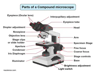
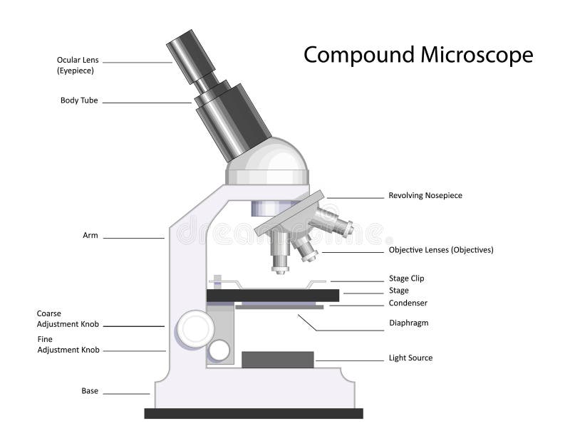
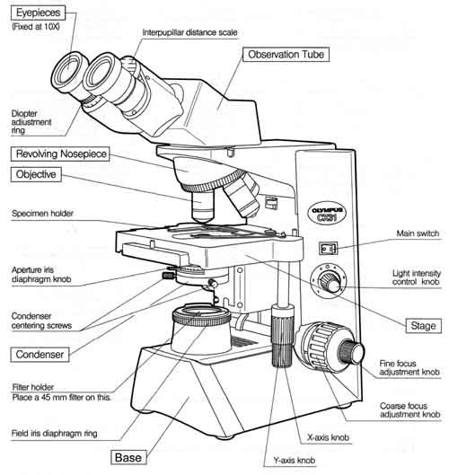
Compound Microscope Parts, Functions, and Labeled Diagram Compound Microscope Definitions for Labels. Eyepiece (ocular lens) with or without Pointer: The part that is looked through at the top of the compound microscope. Eyepieces typically have a magnification between 5x & 30x. Monocular or Binocular Head: Structural support that holds & connects the eyepieces to the objective lenses.
Microscope - Drawing - Worksheet - Estrutura Do Feijão, HD ...
Microscope Parts, Function, & Labeled Diagram - slidingmotion Microscope parts labeled diagram gives us all the information about its parts and their position in the microscope. Microscope Parts Labeled Diagram The principle of the Microscope gives you an exact reason to use it. It works on the 3 principles. Magnification Resolving Power Numerical Aperture. Parts of Microscope Head Base Arm Eyepiece Lens

compound light microscope - Clip Art Library
Microscope Diagram Labeled, Unlabeled and Blank | Parts of a Microscope ... Description Worksheet identifying the parts of the compound light microscope. Answer key: 1. Body tube 2. Revolving nosepiece 3. Low power objective 4. Medium power objective 5. High power objective 6. Stage clips 7. Diaphragm 8. Light source 9. Eyepiece 10. Arm 11. Stage 12. Coarse adjustment knob 13. Fine adjustment knob 14. Base S

Microscopy
Microscope Labeled Pictures, Images and Stock Photos Browse 49 microscope labeled stock photos and images available, or start a new search to explore more stock photos and images. ... Stem and leaves labeled closeup drawings with layers and cells. Educational biology poster. White blood cell types labeled examples educational vector illustration. White blood cell types labeled examples ...
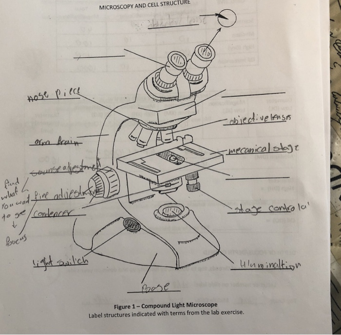
MICROSCOPY AND CELL STRUCTURE (C Figure 1- Compound | Chegg.com
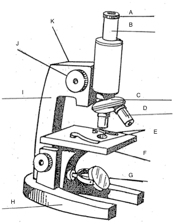
Label Microscope Diagram - EnchantedLearning.com Using the terms listed below, label the microscope diagram. arm - this attaches the eyepiece and body tube to the base. base - this supports the microscope. body tube - the tube that supports the eyepiece. coarse focus adjustment - a knob that makes large adjustments to the focus. diaphragm - an adjustable opening under the stage, allowing ...

Microscope - diagram Tom Butler | Science skills, Microscope ...
Microscope With Labels Clip Art at Clker.com Download Clker's Microscope With Labels clip art and related images now. Multiple sizes and related images are all free on Clker.com.
Microscope, Microscope Parts, Labeled Diagram, and Functions
Labeling the Parts of the Microscope Labeling the Parts of the Microscope This activity has been designed for use in homes and schools. Each microscope layout (both blank and the version with answers) are available as PDF downloads. You can view a more in-depth review of each part of the microscope here. Download the Label the Parts of the Microscope PDF printable version here.

Compound Microscope Parts – Labeled Diagram and their ...
Parts of the Microscope with Labeling (also Free Printouts) Parts of the Microscope with Labeling (also Free Printouts) A microscope is one of the invaluable tools in the laboratory setting. It is used to observe things that cannot be seen by the naked eye. Table of Contents 1. Eyepiece 2. Body tube/Head 3. Turret/Nose piece 4. Objective lenses 5. Knobs (fine and coarse) 6. Stage and stage clips 7. Aperture

Light Microscope PNG - light-microscope-e compound-light ...
Microscope, Microscope Parts, Labeled Diagram, and Functions Microscope, Microscope Parts, Labeled Diagram, and Functions What is Microscope? A microscope is a laboratory instrument used to examine objects that are too small to be seen by the naked eye. It is derived from Ancient Greek words and composed of mikrós, "small" and skopeîn,"to look" or "see".

Microscope Diagram Diagram | Quizlet
Simple Microscope - Parts, Functions, Diagram and Labelling Parts of the optical parts are as follows: Mirror - A simple microscope has a plano-convex mirror and its primary function is to focus the surrounding light on the object being examined. Lens - The biconvex lens is placed above the stage and its function is to magnify the size of the object being examined.

Vektor Stok Microscope Diagram Vector Illustration Labeled ...
Microscope Labeling - The Biology Corner Microscope Labeling Microscope Labeling 15. When focusing a specimen, you should always start with the _____________ objective. 16. When using the high power objective, only the _______________ knob should be used. 17. The type of microscope used in most science classes is the ______________ microscope. 18.

Collection Of Free Microscopes Drawing Label Clipart ...
Label the microscope — Science Learning Hub All microscopes share features in common. In this interactive, you can label the different parts of a microscope. Use this with the Microscope parts activity to help students identify and label the main parts of a microscope and then describe their functions. Drag and drop the text labels onto the microscope diagram.
Compound Microscope Parts – Labeled Diagram and their ...
Microscope Drawing Teaching Resources | Teachers Pay Teachers Microscope Practice: Three Assorted Specimens Drawing (Half Sheets) by GEEKS 7 $1.00 PDF Are your students learning how to use a microscope? This GEEKS lab sheet gives students room to draw three specimens. It includes labels for the Specimen Name, Magnification, Observations/ Challenges.

How To Draw A Microscope, Step by Step, Drawing Guide, by ...
Compound Microscope- Definition, Labeled Diagram, Principle, Parts, Uses The optical microscope often referred to as the light microscope, is a type of microscope that uses visible light and a system of lenses to magnify images of small subjects. There are two basic types of optical microscopes: Simple microscopes. Compound microscopes. The term "compound" in compound microscopes refers to the microscope having ...

Simple Microscope - Diagram (Parts labelled), Principle ...
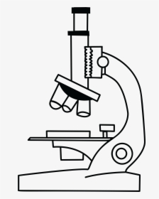
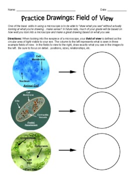
Microscope Drawing: How to Sketch Microscope Slides How to Draw Microscope Slides Organize and orient your field of view: To begin, draw a circle as large as possible with a pencil. An 8.5 x 11-inch piece of paper is good size for beginners. The circle represents what you see through the eyepiece of the microscope. Using thin lines, divide the circle into quarters in order to organize the picture.

Draw a labelled diagram of a compound microscope.
A Study of the Microscope and its Functions With a Labeled Diagram A Study of the Microscope and its Functions With a Labeled Diagram To better understand the structure and function of a microscope, we need to take a look at the labeled microscope diagrams of the compound and electron microscope. These diagrams clearly explain the functioning of the microscopes along with their respective parts.

Microscope With Labels Clip Art at Clker.com - vector clip ...
Microscope- Definition, Parts, Functions, Types, Diagram, Uses

How to Draw a Microscope - Really Easy Drawing Tutorial

microscope drawing with label - Clip Art Library
Microscope Stock Illustrations – 102,378 Microscope Stock ...
Vektor Stok Vector Microscope (Tanpa Royalti) 1209424708 ...

Parts of Stereo Microscope (Dissecting microscope) – labeled ...
![How To Draw A Microscope Step by Step - [12 Easy Phase]](https://easydrawings.net/wp-content/uploads/2021/01/Overview-for-Microscope-drawing.jpg)
How To Draw A Microscope Step by Step - [12 Easy Phase]

Old microscope color sketch engraving vector illustration ...

Cell Drawing Microscope - Binocular Compound Microscope ...

Diagram of traveling microscope setup with implant cast and ...

microscopy how a microscope works magnification calculations ...

How to draw compound of Microscope easily - step by step

Can someone can send me diagram of this compound microscope ...
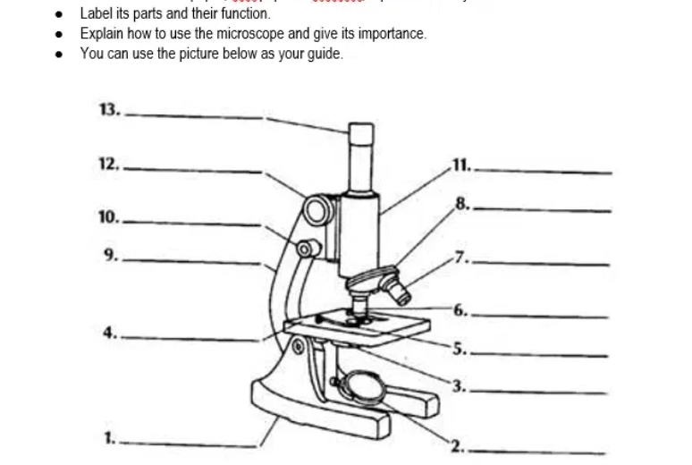
Answered: Label its parts and their function.… | bartleby

Microscope Diagram Labeled, Unlabeled and Blank | Parts of a ...

Biology Notes for A level: #75 Drawings

Course: s4: Biology , Topic: UNIT 3: MICROSCOPY

Diagram of a Compound Microscope

Drawing Microscope Stock Illustrations – 6,450 Drawing ...

Microscope Drawing posted by Samantha Walker

Label Microscope Diagram - EnchantedLearning.com
Microscope | Other Quiz - Quizizz
Untitled Document

Types of Microscopes: Definition, Working Principle, Diagram ...

Label microscope pt.1 Diagram | Quizlet
Microscope Drawing Teaching Resources | Teachers Pay Teachers
Post a Comment for "43 microscope drawing with label"