39 labeled microscope drawing
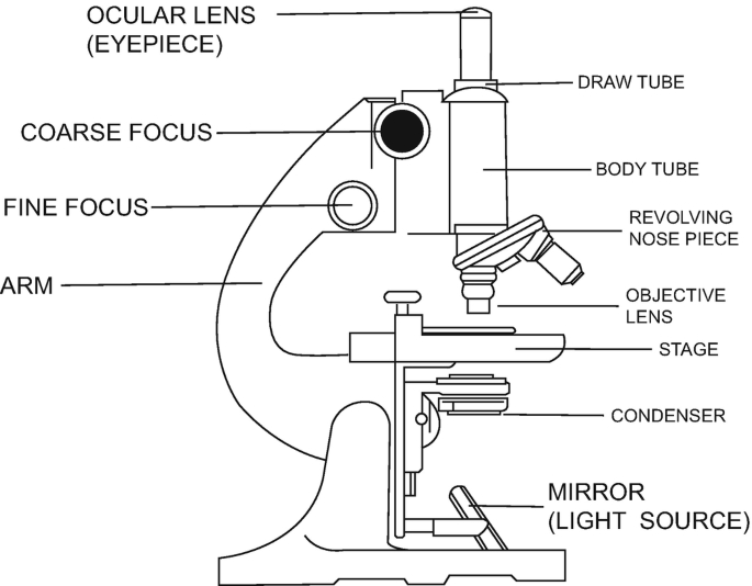
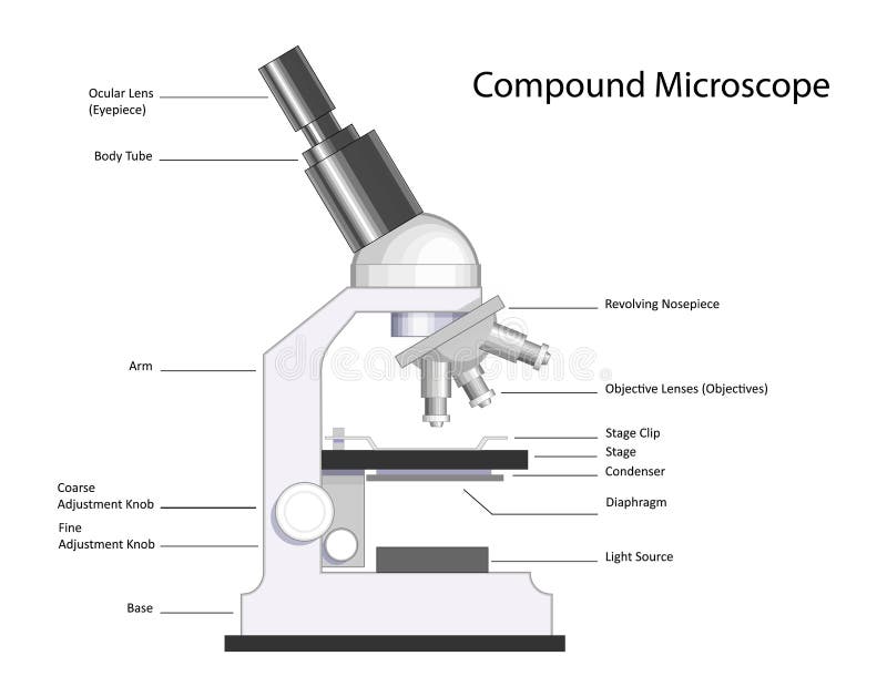
Compound Microscope - Diagram (Parts labelled), Principle and Uses See: Labeled Diagram showing differences between compound and simple microscope parts Structural Components The three structural components include 1. Head This is the upper part of the microscope that houses the optical parts 2. Arm This part connects the head with the base and provides stability to the microscope. Microscope Diagram Labeled, Unlabeled and Blank | Parts of a Microscope ... timvandevall.com Microscope Diagram Labeled, Unlabeled and Blank | Parts of a Microscope Print a microscope diagram, microscope worksheet, or practice microscope quiz in order to learn all the parts of a microscope. Tim's Printables 39k followers More information Microscope Diagram Find this Pin and more on Science Printables by Tim's Printables.
Ray - math word definition - Math Open Reference It has zero width. If you draw a ray with a pencil, examination with a microscope would show that the pencil mark has a measurable width. The pencil line is just a way to illustrate the idea on paper. In geometry however, a ray has no width. A ray has no measurable length, because it goes on forever in one direction. Drawing a ray

Labeled microscope drawing
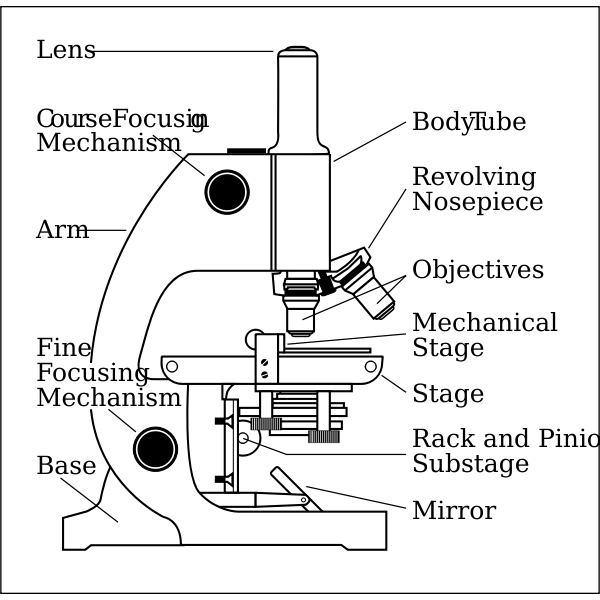
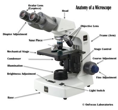
Parts of a microscope with functions and labeled diagram - Microbe Notes Figure: Diagram of parts of a microscope There are three structural parts of the microscope i.e. head, base, and arm. Head - This is also known as the body. It carries the optical parts in the upper part of the microscope. Base - It acts as microscopes support. It also carries microscopic illuminators. Compound Microscope Parts, Functions, and Labeled Diagram Compound Microscope Parts, Functions, and Labeled Diagram Parts of a Compound Microscope Each part of the compound microscope serves its own unique function, with each being important to the function of the scope as a whole. Microscope Parts, Function, & Labeled Diagram - slidingmotion Microscope parts labeled diagram gives us all the information about its parts and their position in the microscope. Microscope Parts Labeled Diagram The principle of the Microscope gives you an exact reason to use it. It works on the 3 principles. Magnification Resolving Power Numerical Aperture. Parts of Microscope Head Base Arm Eyepiece Lens
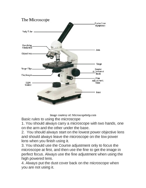
Labeled microscope drawing. Compound Microscope- Definition, Labeled Diagram, Principle ... Apr 03, 2022 · A compound microscope is of great use in pathology labs so as to identify diseases. Various crime cases are detected and solved by drawing out human cells and examining them under the microscope in forensic laboratories. The presence or absence of minerals and the presence of metals can be identified using compound microscopes. Microscope World | Shop Microscopes For Every Application Labeling the Parts of the Microscope. This activity has been designed for use in homes and schools. Each microscope layout (both blank and the version with answers) are available as PDF downloads. You can view a more in-depth review of each part of the microscope here. Microscope labeled diagram - SlideShare Microscope labeled diagram 1. The Microscope Image courtesy of: Microscopehelp.com Basic rules to using the microscope 1. You should always carry a microscope with two hands, one on the arm and the other under the base. 2. You should always start on the lowest power objective lens and should always leave the microscope on the low power lens ... How To Draw A Microscope - YouTube Today, we're learning how to draw a cool microscope!👩🎨 JOIN OUR ART HUB MEMBERSHIP! VISIT 🎨 VISIT OUR AMAZON ART SUPPLY S...
Interactive Bacteria Cell Model - CELLS alive Appendages. Bacteria may have the following appendages. Pili, Fimbriae: These hollow, hairlike structures made of protein allow bacteria to attach to other cells.A specialized pilus, the sex pilus, allows the transfer of plasmid DNA from one bacterial cell to another. How to Sketch a Microscope Slide - Identifying and Sketching Cell ... How to Sketch a Microscope Slide Identifying Cell Structures and Adding Dynamic Elements. Learning how to sketch a microscope slide requires an open-mind, patience and a willingness to learn the basic drawing principles of perspective, size, shape and negative space.. Sketching specimens will provide you with a better understanding, as you study the intricacies of the image you see through the ... Microscope Types (with labeled diagrams) and Functions Simple microscope labeled diagram Simple microscope functions It is used in industrial applications like: Watchmakers to assemble watches Cloth industry to count the number of threads or fibers in a cloth Jewelers to examine the finer parts of jewelry Miniature artists to examine and build their work Also used to inspect finer details on products PDF Label parts of the Microscope: Answers Label parts of the Microscope: Answers Coarse Focus Fine Focus Eyepiece Arm Rack Stop Stage Clip . Created Date: 20150715115425Z ...
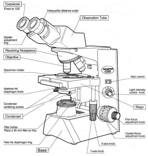
(PDF) Industrial Training Report - Academia.edu 9 CHAPTER TWO 2.1 GENERAL LABORATORY EQUIPMENTS THE LIGHT MICROSCOPE The microscope employs a hollow, extremely intense cone of light concentrated on the specimen. The field of view of the objective lens lies in the hollow, dark portion of the cone and picks up only scattered light from the object. Lab Report Template – Easy Peasy All-in-One High School * All tables, graphs and charts should be labeled appropriately (X and Y axis) Conclusions: * Accept or reject your hypothesis. * EXPLAIN why you accepted or rejected your hypothesis using data from the lab. * Include a summary of the data – averages, highest, lowest..etc to help the reader understand your results. Cells - University of Utah Virtual Microscope Learn how cells work together in tissues, organs, and organ systems. In 1665, Robert Hooke coined the term cell to describe the structures he could see in cork with some of the first microscopes. Microscope, Microscope Parts, Labeled Diagram, and Functions Multiply the magnification of the eyepiece (ocular lens) by the magnification of the objective lens in use to calculate the total magnification of any object viewed under the microscope. This can be demonstrated using the formula. Total magnification = ocular lens x objective lens
Label the microscope — Science Learning Hub
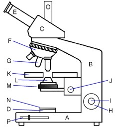
Microscope Labeling - The Biology Corner Microscope Labeling. This simple worksheet pairs with a lesson on the light microscope, where beginning biology students learn the parts of the light microscope and the steps needed to focus a slide under high power. The labeling worksheet could be used as a quiz or as part of direct instruction where students label the microscope as you go ...

BIOLOGY FROM 1 | EQUIPMENTS USED FOR OBSERVATION | Cours ...
Microscope Labeled Pictures, Images and Stock Photos Browse 49 microscope labeled stock photos and images available, or start a new search to explore more stock photos and images. Newest results Fluorescent Imaging immunofluorescence of cancer cells growing... Microscope diagram vector illustration. Labeled zoom instrument... Microscope diagram vector illustration.

Microscope Diagram Labeled, Unlabeled and Blank | Parts of a ...
18,701 Microscope drawing Images, Stock Photos & Vectors - Shutterstock Find Microscope drawing stock images in HD and millions of other royalty-free stock photos, illustrations and vectors in the Shutterstock collection. Thousands of new, high-quality pictures added every day.

How To Draw A Microscope, Step by Step, Drawing Guide, by ...
A Study of the Microscope and its Functions With a Labeled Diagram ... To better understand the structure and function of a microscope, we need to take a look at the labeled microscope diagrams of the compound and electron microscope. These diagrams clearly explain the functioning of the microscopes along with their respective parts. Man's curiosity has led to great inventions. The microscope is one of them.
Microscope side vector drawing with parts labelled | Free SVG
Microscope Drawing Easy with Label - YouTube Microscope Drawing Easy with Label 886 views Apr 13, 2020 In this video I go over a microscope drawing that is easy with label. There is a blank copy at the end of the video to review on your own....

Microscope Labeling
Compound Microscope Parts - Labeled Diagram and their Functions Labeled diagram of a compound microscope Major structural parts of a compound microscope There are three major structural parts of a compound microscope. The head includes the upper part of the microscope, which houses the most critical optical components, and the eyepiece tube of the microscope.
Untitled Document
Microscope Labeling - The Biology Corner Students label the parts of the microscope in this photo of a basic laboratory light microscope. Can be used for practice or as a quiz. ... 20. A microscope has an ocular objective of 10x and a high power objective of 50x, what is the microscope's total magnification? _____

Living Environment Course
Microscopy - Wikipedia The field of microscopy (optical microscopy) dates back to at least the 17th-century.Earlier microscopes, single lens magnifying glasses with limited magnification, date at least as far back as the wide spread use of lenses in eyeglasses in the 13th century but more advanced compound microscopes first appeared in Europe around 1620 The earliest practitioners of microscopy include Galileo ...
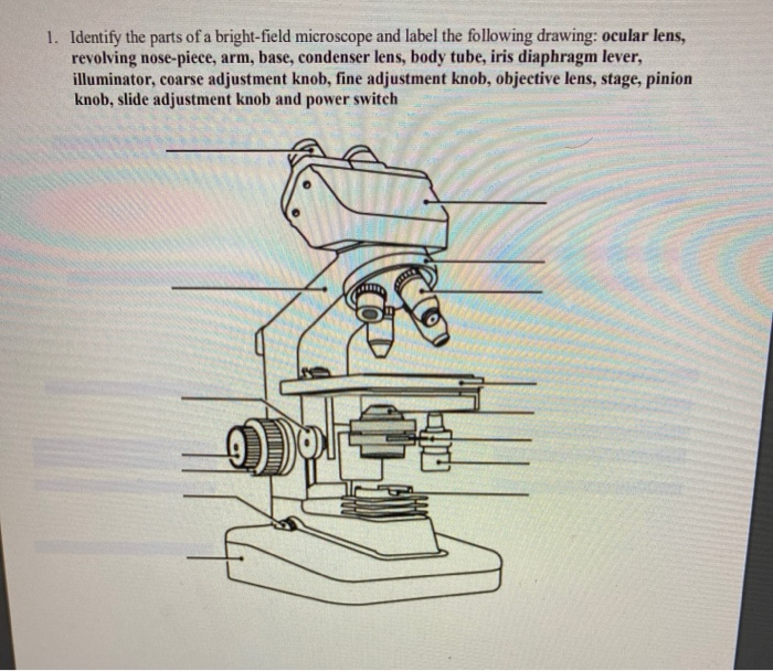
Solved 7. The Microscope 1. In a compound microscope: a. The ...
Labelled Diagram of Compound Microscope The below mentioned article provides a labelled diagram of compound microscope. Part # 1. The Stand: The stand is made up of a heavy foot which carries a curved inclinable limb or arm bearing the body tube. The foot is generally horse shoe-shaped structure (Fig. 2) which rests on table top or any other surface on which the microscope in kept.
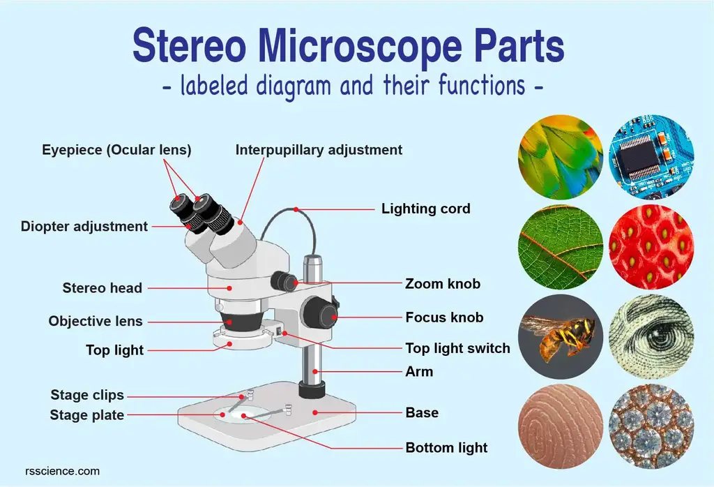
Parts of Stereo Microscope (Dissecting microscope) – labeled ...
Parts of Stereo Microscope (Dissecting microscope) - labeled diagram ... Stereo microscopes (also called Dissecting microscope) are branched out from other light microscopes for the application of viewing "3D" objects. These include substantial specimens, such as insects, feathers, leaves, rocks, sand grains, gems, coins, and stamps, etc. Functionally, a stereo microscope is like a powerful magnifying glass.

Types of Microscopes: Definition, Working Principle, Diagram ...
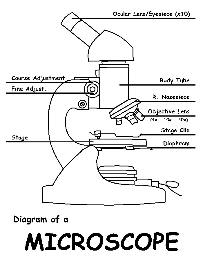
Label Microscope Diagram - EnchantedLearning.com Using the terms listed below, label the microscope diagram. arm - this attaches the eyepiece and body tube to the base. base - this supports the microscope. body tube - the tube that supports the eyepiece. coarse focus adjustment - a knob that makes large adjustments to the focus. diaphragm - an adjustable opening under the stage, allowing ...
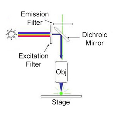
Fluorescence Microscopy - Explanation and Labelled Images ...
Label Microscope Diagram - EnchantedLearning.com Using the terms listed below, label the microscope diagram. arm - this attaches the eyepiece and body tube to the base. base - this supports the microscope. body tube - the tube that supports the eyepiece. coarse focus adjustment - a knob that makes large adjustments to the focus. diaphragm - an adjustable opening under the stage, allowing ...

How to Draw a Microscope - VERY EASY
label microscope diagram | Charts | Microscope, Anatomy bones, Diagram ... Feb 26, 2020 - Microscope Diagram - Microscope - Microscope Parts - Diagram of a Microscope - Parts of a microscope diagram - Electron Microscope - Microscope Magnification - Microscope diagrams. Light microscope, optical microscope diagrams. Label microscope diagram. Microscope labeled diagram. Microscope lens.

Compound Microscope Parts, Diagram Definition, Application ...
Microscope Parts and Functions Most specimens are mounted on slides, flat rectangles of thin glass. The specimen is placed on the glass and a cover slip is placed over the specimen. This allows the slide to be easily inserted or removed from the microscope. It also allows the specimen to be labeled, transported, and stored without damage.

Microscope Drawing Worksheet | Clipart library - Free Clipart ...
Label the microscope — Science Learning Hub All microscopes share features in common. In this interactive, you can label the different parts of a microscope. Use this with the Microscope parts activity to help students identify and label the main parts of a microscope and then describe their functions. Drag and drop the text labels onto the microscope diagram.

Draw a well labelled diagram of a microscope. - Brainly.in
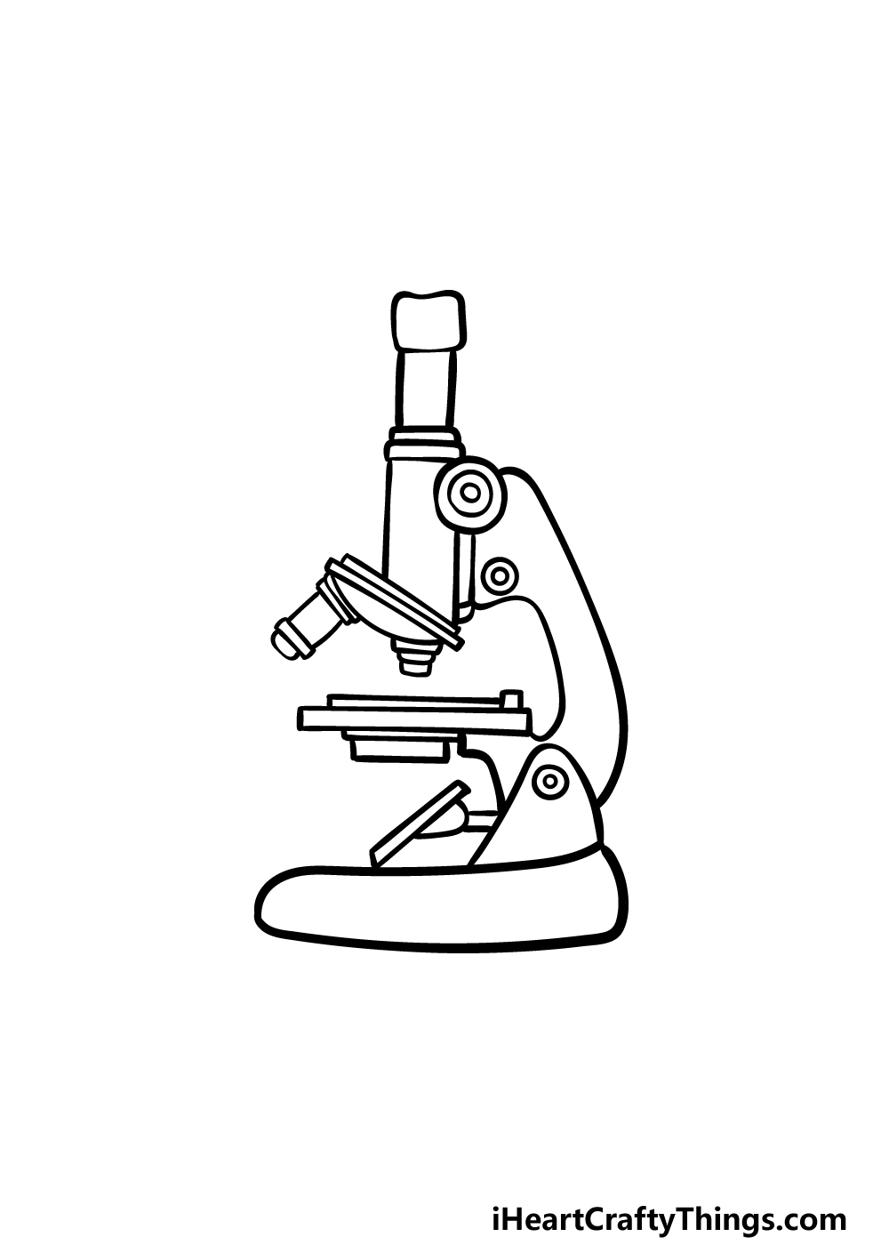
Drawing Of A Microscope And Label - Warehouse of Ideas Here presented 54+ microscope drawing and label images for free to download, print or share. Title Is Informative, Centered, And Larger Than Other Text. How to draw a microscope and label. Compound microscopes have furthered medical research, helped to solve crimes, and they have repeatedly proven invaluable in unlocking the secrets of the.
Microscope Parts and Functions
Parts of the Microscope with Labeling (also Free Printouts) Parts of the Microscope with Labeling (also Free Printouts) By Editorial Team March 7, 2022 A microscope is one of the invaluable tools in the laboratory setting. It is used to observe things that cannot be seen by the naked eye. Table of Contents 1. Eyepiece 2. Body tube/Head 3. Turret/Nose piece 4. Objective lenses 5. Knobs (fine and coarse) 6.

Tsetse biology, systematics and distribution, techniques
Label the Microscope Diagram | Download Scientific Diagram - ResearchGate Download scientific diagram | Label the Microscope Diagram from publication: Laboratory Exercises in Microbiology: Discovering the Unseen World through Hands-on Investigation | Microbiology ...
Free Microscope Drawing, Download Free Microscope Drawing png ...
Microscope Parts, Function, & Labeled Diagram - slidingmotion Microscope parts labeled diagram gives us all the information about its parts and their position in the microscope. Microscope Parts Labeled Diagram The principle of the Microscope gives you an exact reason to use it. It works on the 3 principles. Magnification Resolving Power Numerical Aperture. Parts of Microscope Head Base Arm Eyepiece Lens

Free Microscope Drawing, Download Free Microscope Drawing png ...
Compound Microscope Parts, Functions, and Labeled Diagram Compound Microscope Parts, Functions, and Labeled Diagram Parts of a Compound Microscope Each part of the compound microscope serves its own unique function, with each being important to the function of the scope as a whole.
Free Microscope Drawing, Download Free Microscope Drawing png ...
Parts of a microscope with functions and labeled diagram - Microbe Notes Figure: Diagram of parts of a microscope There are three structural parts of the microscope i.e. head, base, and arm. Head - This is also known as the body. It carries the optical parts in the upper part of the microscope. Base - It acts as microscopes support. It also carries microscopic illuminators.

Compound Microscope- Definition, Labeled Diagram, Principle ...

Label the microscope — Science Learning Hub
Exercise 1: Using a Compound Microscope | SpringerLink
Microscope Drawing - How To Draw A Microscope Step By Step

Microscope - Label - Part 2 Diagram | Quizlet

Bagaimana Menggambar Mikroskop
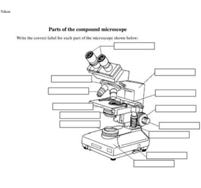
Solved Nikon Parts of the compound microscope Write the ...
Microscope Stock Illustrations – 104,693 Microscope Stock ...
![How To Draw A Microscope Step by Step - [12 Easy Phase]](https://easydrawings.net/wp-content/uploads/2021/01/Overview-for-Microscope-drawing.jpg)
How To Draw A Microscope Step by Step - [12 Easy Phase]

Diagram of a Compound Microscope

Microscope Labeling Diagram | Quizlet
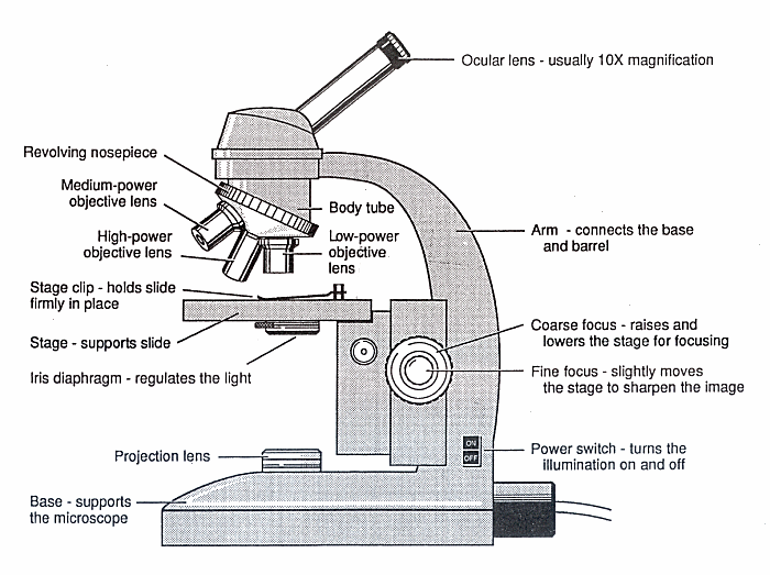
Diagram of a Microscope - Guide to using a microscope

Microscope - diagram Tom Butler | Science skills, Science ...

Simple Microscope - Parts, Functions, Diagram and Labelling ...
MICROSCOPE LAB
Glossary of terms used in microscopy – Quekett Microscopical Club

Light Microscope PNG - light-microscope-e compound-light ...

label the parts of the compound microscope - Brainly.ph
Post a Comment for "39 labeled microscope drawing"