44 unlabeled sheep brain
sheep brain labeling Quiz - PurposeGames.com sheep brain labeling by natalee1438 7,341 plays 22 questions About a minute 7 too few (you: not rated) Language English Tries Unlimited [?] Last Played February 22, 2022 - 12:00 am There is a printable worksheet available for download here so you can take the quiz with pen and paper. Remaining 0 Correct 0 Wrong 0 Press play! 0% 12:00.0 Highscores 2,882 Labeled Brain Anatomy Images, Stock Photos & Vectors - Shutterstock Find Labeled Brain Anatomy stock images in HD and millions of other royalty-free stock photos, illustrations and vectors in the Shutterstock collection. Thousands of new, high-quality pictures added every day.
ARC Sheep Brain Practical - Anki Flashcards - Ben Millam they are not labeled, structures are identified only with a dot (approximating a pin) · green dots indicate the card is a nerve (nerves may be ambiguous on some ...

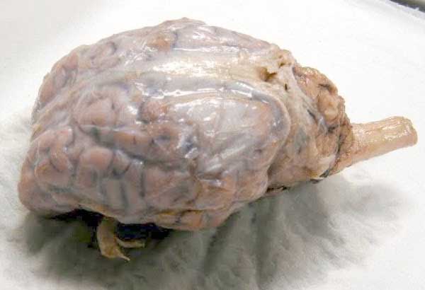
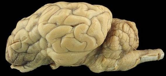
Unlabeled sheep brain
Sheep Brain Labeling (part 1) Quiz - By dilatory - Sporcle Sheep Brain Labeling (part 1) Quiz - By dilatory Science sheep QUIZ LAB SUBMISSION Random Science or sheep Quiz Sheep Brain Labeling (part 1) Can you name the Sheep Brain parts? By dilatory Plays - /5 - RATE QUIZ MORE INFO Classic Best Score? Go Orange. hide this ad PLAY QUIZ Score 0/25 Timer 10:00 Recently Published The Sheep Brain Atlas - Michigan State University The Sheep Brain Atlas John I. Johnson, Keith D. Sudheimer, Kristina K. Davis, Garrett M. Kerndt, and Brian M. Winn Radiology Department, Neuroscience Program ,and Communications Technology Laboratory, Michigan State University, East Lansing, MI Please let us know if you are using any of these atlases or images for particular classes. Sheep Brain Dissection labeled Diagram | Quizlet Sheep Brain Dissection labeled + − Learn Test Match Created by AllieKlinger Terms in this set (8) Corpus Collosum ... Lateral Ventricle ... Fornix ... Hypothalamus ... Cerebral Aqueduct ... Central Canal ... Inferior Collicuious ... Transverse Fissure ... Students also viewed Sheep Brain Dissection, Sheep Brain Diss… 65 terms sydneypfleiger
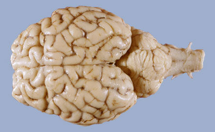
Unlabeled sheep brain. Sheep Brain - Dorsal View - University of Minnesota Dorsal view of sheep brain with the cerebellum and caudal cerebrum removed. The rostral colliculus (large arrow label) and the caudal colliculus (small arrow label) together form the tectum of the midbrain.. Also labeled are the pineal body (green), the caudate nucleus (1), the floor of the fourth ventricle (white and pink) and cerebellar peduncles (blue = rostral, red = middle, and yellow ... Sheep brain Flashcards | Quizlet Identification of structures observed during sheep brain dissection. Terms in this set (29) dura mater Identify the covering. cerebrum Identify the major brain region. cerebellum Identify the major brain region. olfactory bulb Identify the tip. optic nerve Identify the nerve by name. optic chiasma Identify the "x". optic chiasma Kami Export - Sheep Brain unlabeled to review for quiz.docx.pdf View Kami Export - Sheep Brain unlabeled to review for quiz.docx.pdf from SCIENCE N/A at Morristown-Beard School. Sheep Brain Labeling Know for Quiz! Name_ sulcus gyrus cerebrum cerebral Sheep Brain Instructions - University of Scranton The Sheep Brain Dissection Guide Uses Frames And It May Not Be Compatible With All Web Browsers!! You have several options when proceeding through this guide. As you progress, you will see the image of the brain being dissected in this window. ... This button allows you to toggle between labeled and unlabeled images. On many of the images you ...
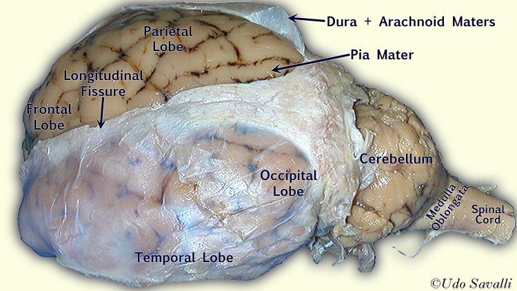
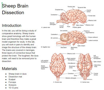
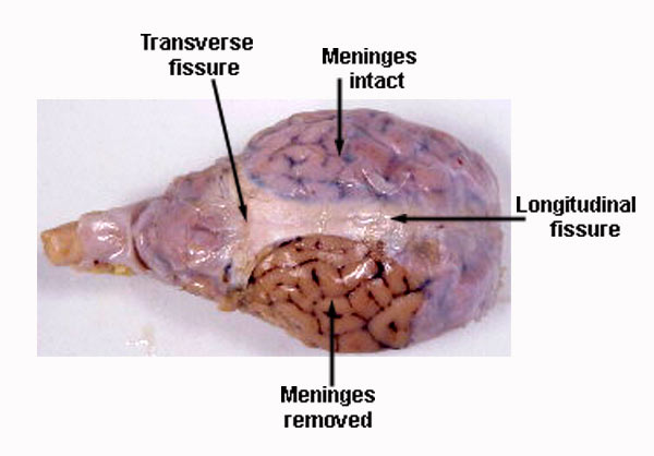
Sheep Brain Label | Dissection, Brain anatomy, Human brain diagram Sheep Brain Label A drawing of the brain with the parts unlabeled. Students can practice naming the parts of the brain, then check their answers with the provided key. Biologycorner 18k followers More information unlabeled brain Nervous System Lesson Nervous System Anatomy Human Brain Diagram Brain Gym For Kids Brain Anatomy And Function Comparative Neuropathology of Ovine Enterotoxemia Produced by ... Clostridium perfringens type D causes enterotoxemia in sheep and goats. ... nohistochemical markers to further characterize the brain lesions induced by ETX ... Sheep Brain Neuroanatomy Online Self-Test | KPU.ca - Kwantlen ... Sheep Brain Neuroanatomy Online Self-Test Use each diagram as a reference, and selected the correct answer for each lettered structure. You may find it useful to open the diagrams in a separate window to review while answering each question. Dorsal Surface Dorsal Surface A * Occipital Lobe Temporal Lobe Cerebellum Parietal Lobe Dorsal Surface B * Sheep Brain Dissection with Labeled Images - The Biology Corner 1. The sheep brain is enclosed in a tough outer covering called the dura mater. You can still see some structures on the brain before you remove the dura mater. Take special note of the pituitary gland and the optic chiasma. These two structures will likely be pulled off when you remove the dura mater. Brain with Dura Mater Intact
Sheep Brain Dissection Guide - The Biology Corner The sheep brain is quite similar to the human brain except for proportion. The sheep has a smaller cerebrum. Also the sheep brain is oriented anterior to posterior whereas the human brain is superior to inferior. 1. The tough outer covering of the sheep brain is the dura mater, one of three meninges (membranes) that cover the brain. PDF Sheep Brain Practical Study Guide - auburn.k12.il.us Sheep Brain Practical Study Guide. Dura Mater. Olfactory Bulb Pituitary Gland Dura Mater Optic Chiasm. Corpus Callosum Longitudinal Fissure Lateral Ventricle Gray Matter White Matter. Arbor Vitae "Tree of Life" Cerebellum "Little Brain" ... Sheep Brain - Ventral View - University of Minnesota Sheep Brain - Ventral View Ventral view of a sheep brain. The optic chiasm (green pic) marks the rostral end of the hypothalamus ( optic nerves are rostral and optic tracts are caudal to the chiasm). Mamillary bodies (red) mark the caudal end of the hypothalamus. Between these, the orange pic is in the lumen of the pituitary stalk (infundibulum). Sheep Brain - Anatomy Corner ... dissection of the sheep's brain, some structures have been labeled. superior colliculus labeled · brain, lateral view · labeled brain · unlabeled brain ...
sheep brain dissection labeling ex. 14 Diagram | Quizlet
Unlabeled | LGBTQIA+ Wiki | Fandom Unlabeled, also known as "no label" or "non-labeled", is a term used by individuals who do not wish to label their identity with more specific terms, such as lesbian, bisexual, agender, and so on. People can have many personal reasons for wanting to forego labeling themselves, such as feeling that current labels do not fit how they feel, or just not wanting to label themselves for the time ...
Lab 12 - The Brain and Cranial Nerves
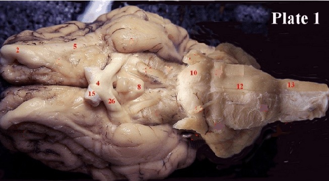
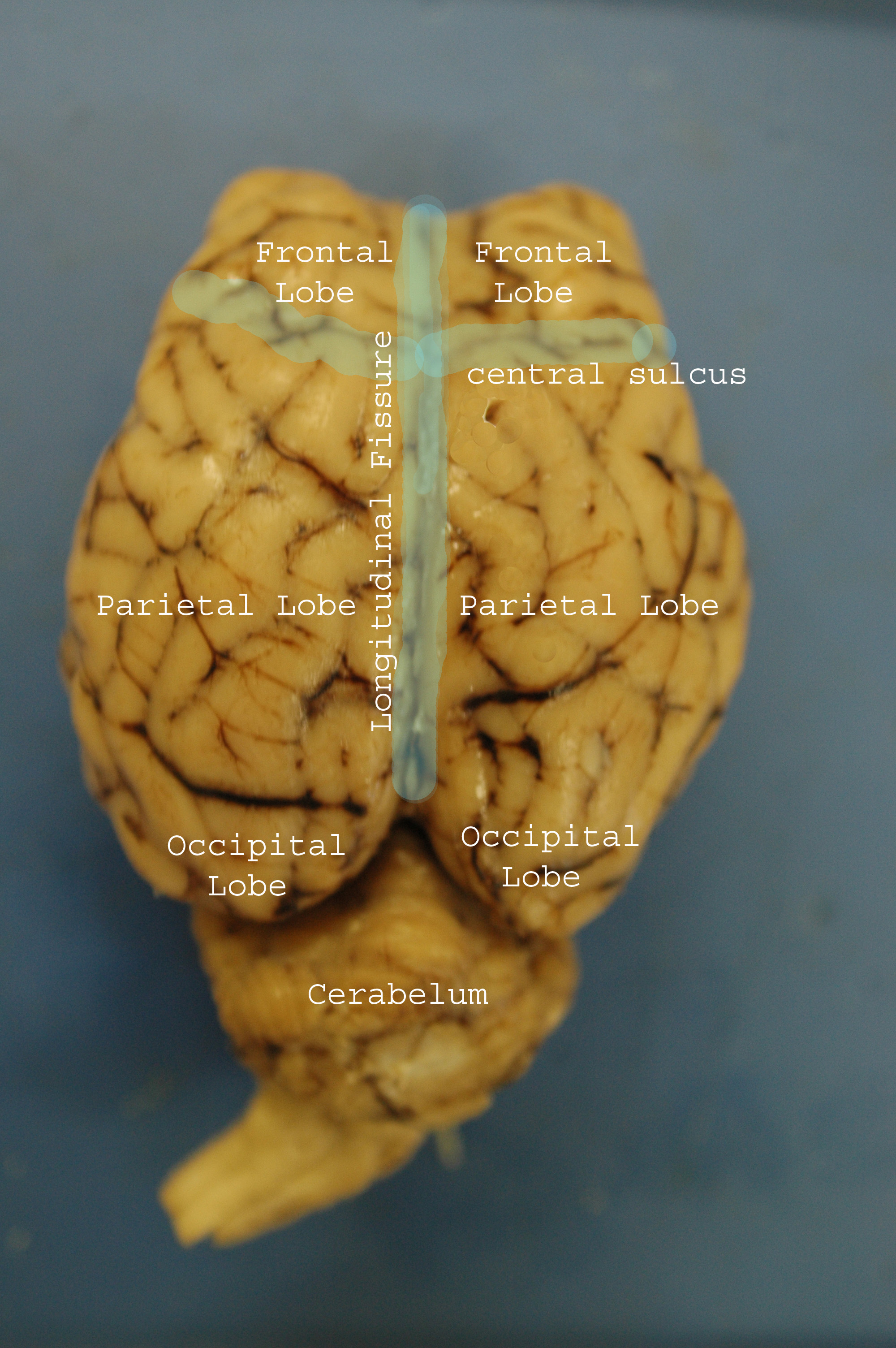
Practice Lab Practical on the Sheep Brain - PGCC Practice Lab Practical on the Sheep Brain In the above picture: Identify the lobe labeled 1. Look here for the answer Frontal Identify the lobe labeled 2. Look here for the answer Temporal Identify the lobe labeled 3. Look here for the answer Parietal Identify the structure labeled 4. Look here for the answer Vermis

Close up of a sheep brain cut in half Stock Photo - Alamy
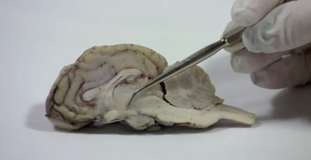
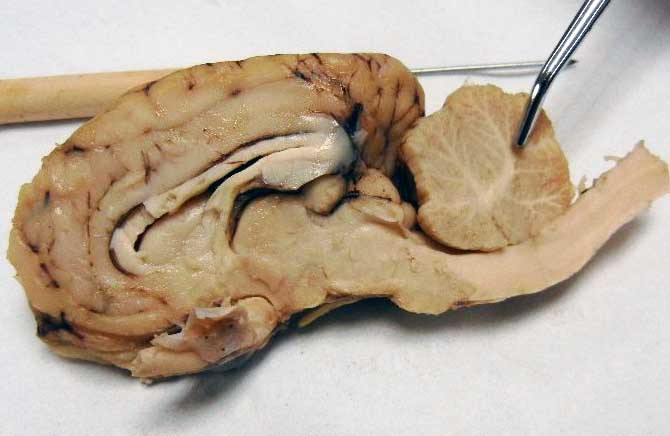
DISSECTION OF THE SHEEP'S BRAIN While not labeled see if you can see the corpus callosum and the lateral ventricles. Hippocampus. Lateral Geniculate. Nucleus. Medial Geniculate. Nucleus.
Sheep Brain Dissection Project Guide | HST Learning Center
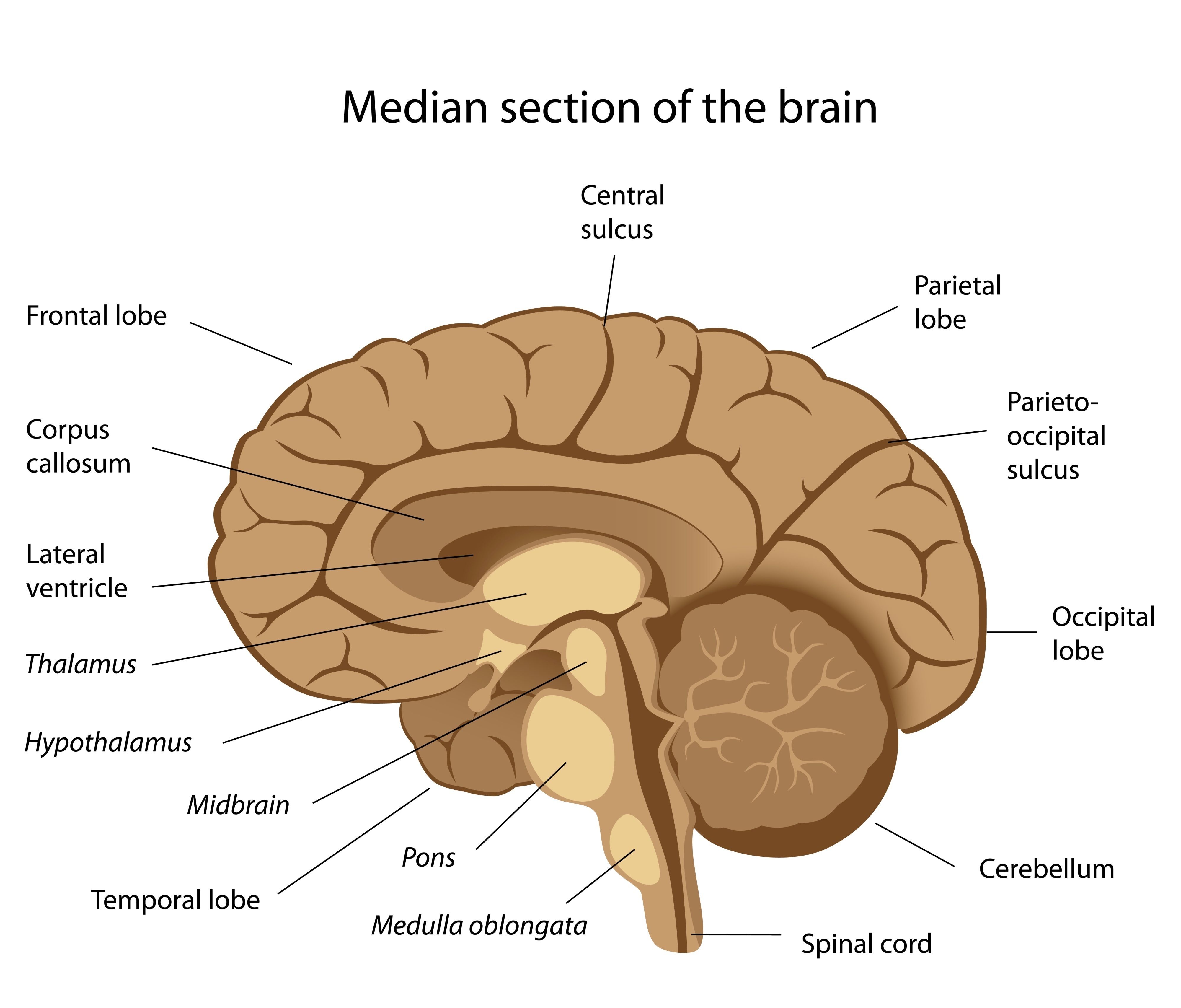
Sheep Brain Anatomy with Labeled Diagram The sheep brain anatomy consists of 3 major parts - prosencephalon (forebrain), mesencephalon (midbrain), and rhombencephalon (hindbrain). These 3 main parts of the sheep brain again divide into specific segments. There are also 5 different lobes in the sheep brain structure - frontal, parietal, occipital, temporal, and limbic area.
Sheep Neuroanatomy Lab- Labeling Worksheet Figure 1: Dorsal view
Unlabeled Sheep Brain Dissection Images and Link (1).pptx BIOL BIOL MISC Unlabeled Sheep Brain Dissection Images and Link (1).pptx - http:/classroom.sdmesa.edu/anatomy/ModelPages/sheep_brain.htm Unlabeled Sheep Brain Dissection Images and Link (1).pptx -... School University of Pennsylvania Course Title BIOL MISC Uploaded By seperry215yahoo.com Pages 9 This preview shows page 1 - 2 out of 9 pages.
BIO201-Sheep Brain
NERVOUS SYSTEM - SHEEP BRAIN IMAGES - sdmesa.edu Sheep Brain Images San Diego Mesa College offers various certificates and degrees to prepare students for careers relating to Biology. skip to main content Class Schedule MySDCCD Canvas Faculty/Staff Alumni Newsroom Calendar Directory Map Library Admissions GIVE TO MESA About
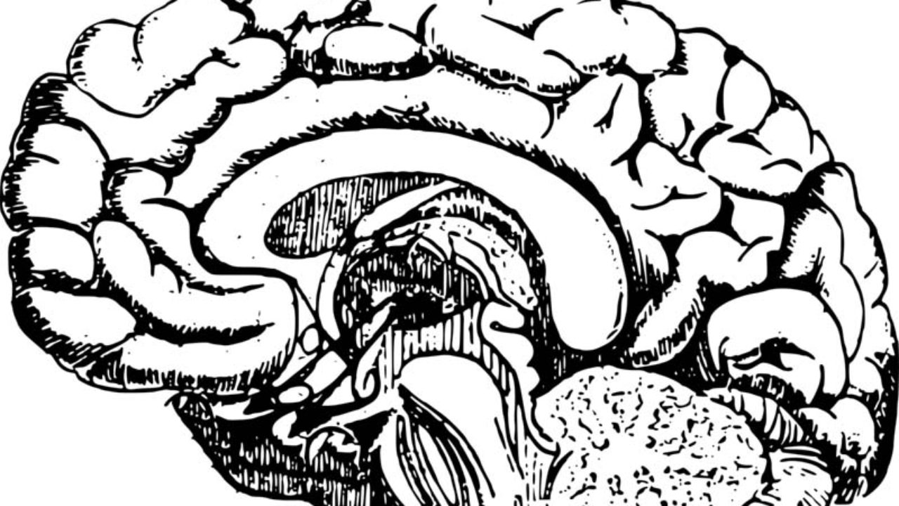
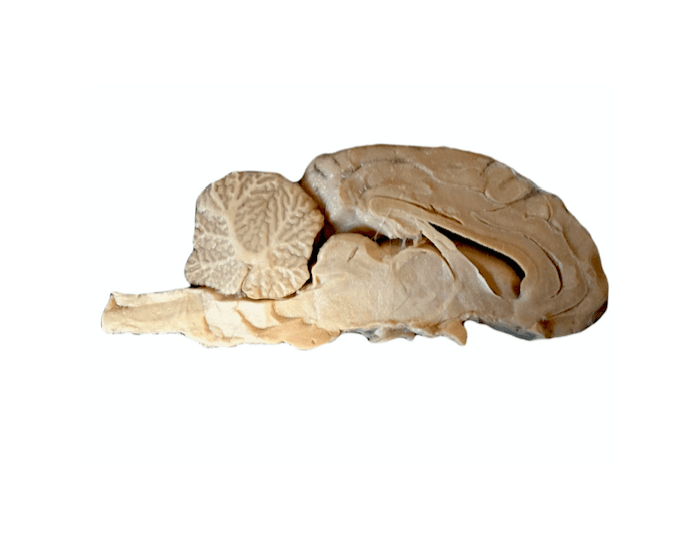
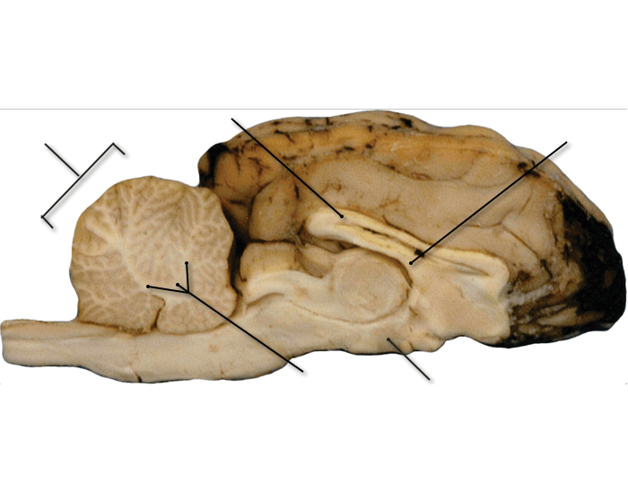
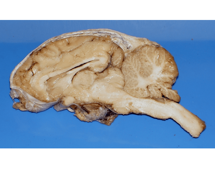
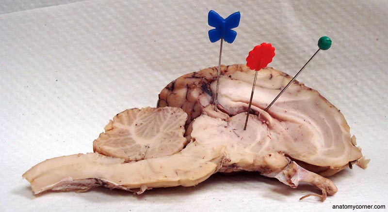
Midsagittal Section
BIO201-Sheep Brain BIO201-Sheep Brain Structures of the preserved sheep brain Return to Unlabeled Photos This page last updated 18 August 2019 by Udo M. Savalli ( dr.udo @ savalli.us) Images and text © Udo M. Savalli. All rights reserved.
Solved Question 4 Identify the parts of the sheep | Chegg.com
Lab 9—Sheep Brain—Labeled - Bluegrass Community and Technical College Lab 9—Sheep Brain—Labeled BIO 137 Virtual Lab 9 The Sheep's Brain Return to: The Unlabeled BrainsLab 9 PageBIO 137 Main Page Be sure to practice identifying the structures using the unlabeled photos. This page created and maintained by Udo M. Savalli. Last updated August 13, 2005.
Sheep Brain sagittal view Quiz
Sheep Brain Dissection Project Guide | HST Learning Center Sheep brains, like other sheep organs, are much smaller than human brains but have similar features. They can be a valuable addition to your study of anatomy. See for yourself what the cerebrum, cerebellum, spinal cord, gray and white matter, and other parts of the brain look like with this sheep brain dissection guide!
Index of /files/OCC_VIDEO/upload/Faculty_Resources/acamilo ...
Label the parts of a Sheep Brain (Midsagittal) - PurposeGames.com This online quiz is called Label the parts of a Sheep Brain (Midsagittal). It was created by member Dr. Smith's BSC 2085L and has 10 questions. It is currently featured in 12 tournaments. This online quiz is called Label the parts of a Sheep Brain (Midsagittal). It was created by member Dr. Smith's BSC 2085L and has 10 questions.

BIO 137 LE 9: Sheep Brain (Midsagittal Section) Diagram | Quizlet
Sheep brain dissection | Human Anatomy and Physiology Lab (BSB ... Sheep brain dissection. Lab Exercises 10-1. The sheep brain is quite similar to the human brain except for proportion. The sheep has a smaller cerebrum.

Dissected sheep's brain, midsagittal | Mrs. Sandy | Flickr
Sheep Brain Dissection Key - Pinterest Nervous System Lesson, Nervous System Anatomy, Human Brain Diagram, Brain Gym For ... of the brain located at biologycorner.com - .
Sheep Brain Dissection Lab
Sheep brain unlabeled - YouTube About Press Copyright Contact us Creators Advertise Developers Terms Privacy Policy & Safety How YouTube works Test new features Press Copyright Contact us Creators ...

Sheep Brain
Labeled Diagrams of the Human Brain You'll Want to Copy Now More than half of the neurons in the brain are found in the cerebellum and only 10% neurons make up the brain. 85% of the brain is cerebral cortex, divided as, 41% frontal lobe, 22% temporal lobe, 19% parietal lobe and 18% occipital lobe. There are 186 million more neurons in the left hemisphere of the brain than the right hemisphere.
Dissection of the Sheep Brain
Sheep Brain Dissection | Human Anatomy Quiz - Quizizz answer choices. temporal lobe. parietal lobe. occipital lobe. frontal lobe. Question 7. 120 seconds. Q. Name the green part of the brain, which is responsible for connecting the left and right sides of the brain, allowing for communication between both hemispheres.
Sheep Brain Dissection
Sheep Brain Dissection labeled Diagram | Quizlet Sheep Brain Dissection labeled + − Learn Test Match Created by AllieKlinger Terms in this set (8) Corpus Collosum ... Lateral Ventricle ... Fornix ... Hypothalamus ... Cerebral Aqueduct ... Central Canal ... Inferior Collicuious ... Transverse Fissure ... Students also viewed Sheep Brain Dissection, Sheep Brain Diss… 65 terms sydneypfleiger

ARC Sheep Brain Practical - Anki Flashcards
The Sheep Brain Atlas - Michigan State University The Sheep Brain Atlas John I. Johnson, Keith D. Sudheimer, Kristina K. Davis, Garrett M. Kerndt, and Brian M. Winn Radiology Department, Neuroscience Program ,and Communications Technology Laboratory, Michigan State University, East Lansing, MI Please let us know if you are using any of these atlases or images for particular classes.
Sheep Brain Dissection with Labeled Images
Sheep Brain Labeling (part 1) Quiz - By dilatory - Sporcle Sheep Brain Labeling (part 1) Quiz - By dilatory Science sheep QUIZ LAB SUBMISSION Random Science or sheep Quiz Sheep Brain Labeling (part 1) Can you name the Sheep Brain parts? By dilatory Plays - /5 - RATE QUIZ MORE INFO Classic Best Score? Go Orange. hide this ad PLAY QUIZ Score 0/25 Timer 10:00 Recently Published
Sheep Brain Practical

brain-sheep-lateral | Brain, unlabeled | biologycorner | Flickr
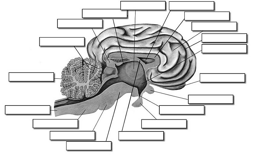
Sheep Brain – Midsagittal Section
Label the parts of a Sheep Brain (Midsagittal) Quiz
Sheep Brain Quiz

Sheep Brain - BNS Lab 2009 EG

Sheep Brain Images
Lab: Sheep Brain Dissection
Sheep Brain Dissection with Labeled Images
DISSECTION OF THE SHEEP'S BRAIN
Sheep Brain Dissection

Welcome to the sheep brain structure review! arachnoid layer ...
hey! i want to die - sheep brain lab practical | Conjunto de ...

Carolina's Perfect Solution® Sheep Brain, Dura Mater Intact, Plain, Pail

Sheep Brain Dissection | Carolina.com

The Brain Lab

brain-sheep-chiasma-1280px | Brain, unlabeled, ventral view ...
University of Scranton Sheep Brain Guide 3.0
Sheep Brain Dissection with Labeled Images
Sheep Brain Label

Sheep Brain

Sheep brain | Atlas of Comparative Vertebrate Anatomy

Sheep Brain Images
midsection of brain labeled - Clip Art Library
Sheep Brain Dissection Bi - BIOLOGY JUNCTION

Median Sagittal Section of Brain
Post a Comment for "44 unlabeled sheep brain"